Surgeons, sports doctors, and biomedical engineers all depend on a deep understanding of how the human body is organized. When a doctor views an MRI scan, they are not just seeing "a leg" or "a brain" — they are mentally mapping layers of systems: muscles, blood vessels, nerves, and more, all stacked and interacting. This layered, nested structure is called a hierarchical organization, and it is a key idea for understanding how complex multicellular organisms work.
Multicellular organisms, like humans, plants, and other animals, are built from many different kinds of cells, each performing specific jobs. The instructions for building and running these cell types are stored in DNA inside almost every cell. While we will not go into molecular or chemical reactions, it is important to know that DNA contains information that leads to different cell types with specialized structures and functions.
These specialized cells are not just scattered randomly. They are organized into a nested hierarchy: cells group into tissues, tissues form organs, organs combine into organ systems, and organ systems interact to form a functioning organism. This hierarchical arrangement allows incredible complexity and coordination while still being built from relatively simple units.
Cell is the basic unit of structure and function in living things.
Tissue is a group of similar cells that work together to perform a specific function.
Organ is a structure made of two or more tissue types working together for a particular task.
Organ system is a group of organs that work together to carry out major body functions such as digestion, circulation, or movement.
In many organisms, this hierarchy is closely tied to inherited information. DNA in each cell contains the instructions for making certain structures (like the tiny hairlike projections on some intestinal cells) that help the organism perform critical functions (like absorbing nutrients). Although we are not examining chemical-level processes, we can see that DNA acts like a high-level "blueprint" for organizing systems within the body.
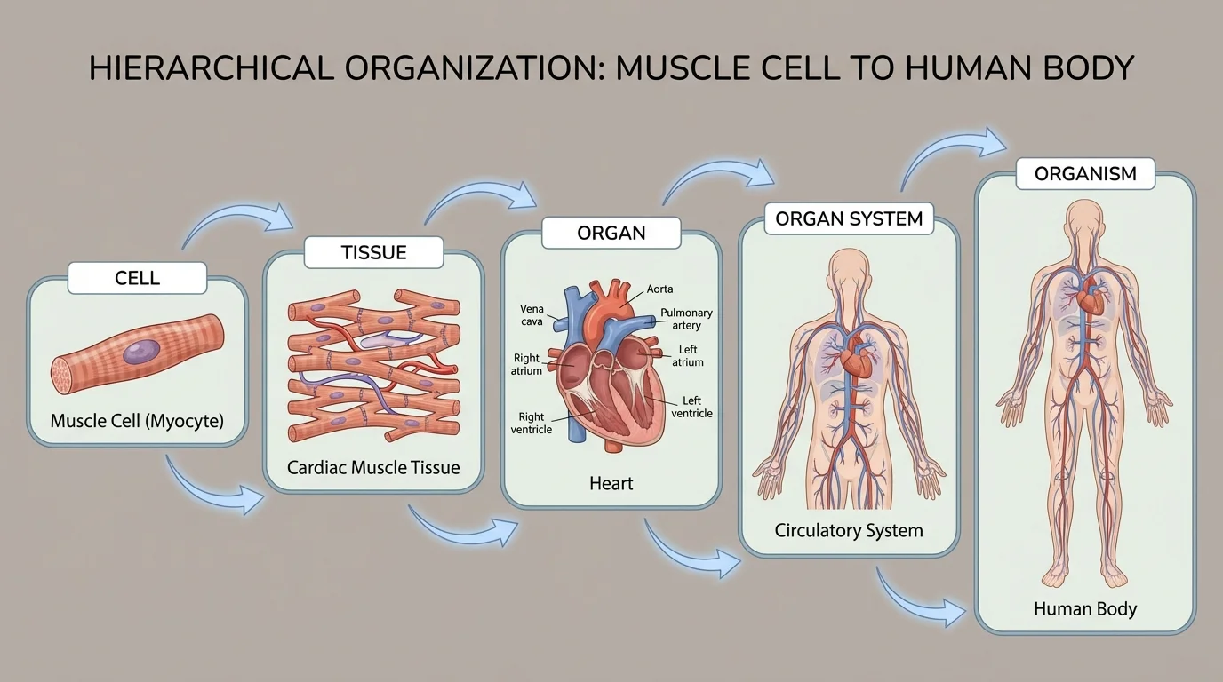
The levels of organization in a typical animal body form a ladder-like hierarchy, as illustrated in [Figure 1]. Each level builds on the one below it and contributes to the function of the one above.
Cells: For example, muscle cells in your heart are long, contractile cells that can shorten and relax. Their structure (with many overlapping fibers) lets them generate force.
Tissues: Similar heart muscle cells group together to form cardiac muscle tissue. This tissue contracts in a coordinated way to squeeze blood out of the heart chambers.
Organs: Cardiac muscle tissue, along with connective tissue, blood vessels, and nerve tissue, form the heart. The heart is an organ with a specific job: pumping blood.
Organ systems: The heart works together with blood vessels (arteries, veins, capillaries) and blood itself to form the circulatory system. This organ system transports materials like oxygen, nutrients, and wastes throughout the body.
Organism: The circulatory system interacts with many other systems (respiratory, digestive, excretory, nervous, etc.) to keep the entire human body — the organism — alive and functioning.
This kind of hierarchy also appears in other systems. In the digestive system, specialized epithelial cells line the stomach; together they form stomach tissue, which builds the stomach organ, which is part of the digestive system, which supports the body's need for energy and building materials.
The key point is that structure and function are linked at every level. The shape, arrangement, and connection of parts at a lower level determine what the next level can do. When you develop a model of a system, you should highlight both the structures at each level and the functions they provide.
Scientists and engineers use the word model in a specific way. A model is not just a picture; it is any representation that helps explain, analyze, or predict how something works. In life science, models of hierarchical systems help us visualize relationships that we cannot see directly.
Common types of models include:
When you develop and use a model of hierarchical organization, you usually want to show at least three things:
Every model has limitations. A 2D diagram might clearly show levels of organization, but it might not show how organs move in 3D space. Recognizing what a model does well and where it is simplified is part of scientific thinking.
When you eat a sandwich, your body has to break it down and absorb its nutrients. The digestive system is a powerful example of how interacting, hierarchical systems provide a specific function: supplying the body with usable nutrients. As food moves along a sequence of organs, each step depends on structures at multiple levels working together, as shown schematically in [Figure 2].
Cells to tissues: The inner surface of your small intestine is lined with specialized epithelial cells. These cells often have tiny projections called villi and microvilli (fingerlike extensions) that greatly increase surface area, allowing more efficient nutrient absorption.
Tissues to organs: In a segment of the small intestine, these epithelial cells combine to form epithelial tissue. Smooth muscle tissue surrounds this lining and contracts rhythmically, pushing food along. Connective tissue and blood vessels support and supply the area. Together, these tissues form the small intestine organ.
Organs to organ system: The small intestine works together with the mouth, esophagus, stomach, liver, pancreas, and large intestine. This collection of organs forms the digestive system. Each organ has specific structures (folds, glands, muscular layers) that contribute to the overall function of breaking down food and absorbing nutrients.
Organ systems to organism: Once nutrients are absorbed into blood vessels in the small intestine, the circulatory system transports them to cells throughout the body. Here we see systems interacting: the digestive system depends on the circulatory system to deliver nutrients, and the circulatory system depends on the digestive system to supply those nutrients in the first place.
If you were to create a model of this hierarchy, you might draw a multi-level diagram: at the bottom, a single intestinal epithelial cell with its microvilli; above that, a cross-section showing epithelial and muscle tissues; above that, the whole small intestine organ; above that, the entire digestive system; and at the top, the whole human. Each level would be labeled with both structure and function (for example, "small intestine: absorbs digested nutrients into blood").
Example: Conceptual model of digestive system hierarchy
Consider a student building a simple conceptual model on paper.
Step 1: List the levels from smallest to largest: cell → tissue → organ → organ system → organism.
Step 2: Choose a specific pathway, such as "intestinal epithelial cell → epithelial tissue → small intestine → digestive system → human body."
Step 3: For each level, write one main function (for example, "intestinal epithelial cell: absorbs glucose and amino acids from digested food").
This model helps the student see how a simple action at the cell level (absorption) contributes all the way up to the organism's survival (getting energy and building materials).
The digestive system also interacts with the nervous system, which regulates the speed of digestion and hunger signals, and with the excretory system, which removes solid wastes. These cross-system interactions show why models often need arrows and labels that connect multiple organ systems, not just a single chain.
Breathing and blood flow are often studied together because they are physically intertwined in the chest. The lungs (respiratory system) and the heart and blood vessels (circulatory system) literally wrap around each other. This close arrangement enables the critical function of gas exchange, as portrayed schematically in [Figure 3].
Cells to tissues in the lungs: The tiny air sacs in your lungs, called alveoli, are lined with very thin epithelial cells. Their thinness and wide surface area allow oxygen to move into nearby blood and carbon dioxide to leave the blood (again, we stay at the structural and system level, not the chemical reaction level).
Tissues to organs in the lungs: These epithelial layers, plus connective tissue and surrounding capillaries (tiny blood vessels), form the lung tissue. Combined on a large scale, they create the lungs, organs that bring air into close contact with blood.
Cells to tissues in the heart and vessels: In the heart, specialized cardiac muscle cells form cardiac muscle tissue, while endothelial cells line the inside of blood vessels. These tissues create a pump (the heart) and tubes (arteries, veins, capillaries) that carry blood.
Organs to interacting organ systems: The respiratory system consists mainly of the lungs and airways, while the circulatory system includes the heart and blood vessels. Together, these systems ensure that oxygen reaches body cells and carbon dioxide is removed.
One way to model their interaction is to trace the journey of a single red blood cell. Starting somewhere in a leg muscle, it travels through veins to the right side of the heart, is pumped into the lungs where it passes by alveoli, picks up oxygen, and returns to the left side of the heart. Then it is pumped out through arteries back to the body. Each stage depends on structures at multiple hierarchical levels.
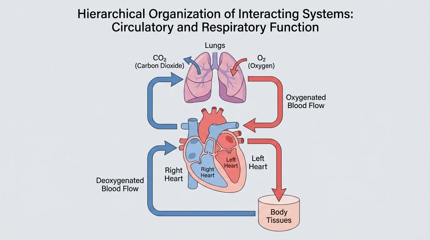
A diagrammatic model, like the one in [Figure 3], is especially useful here. It can show how the heart chambers, blood vessels, and lungs physically connect, and how their coordinated structure produces the overall function of oxygen delivery.
Some diving mammals, like seals, have unusually large blood volumes relative to their body size, allowing their circulatory systems to store more oxygen for long dives. Their hierarchical organization is similar to humans, but certain organs and tissues are adapted to extreme conditions.
Notice that even though the respiratory and circulatory systems are often studied separately in textbooks, in reality their structures are tightly interdependent. Good models make these interactions visible.
Hierarchical organization is not only about structure; it is also about control and regulation. Your body must constantly adjust heart rate, breathing rate, and blood flow based on activity level and environment. The nervous system and endocrine system (hormone system) play major roles in coordinating these changes.
For example, when you run up stairs, sensors in muscles and blood vessels detect changes like increased carbon dioxide levels and muscle activity. Nerve signals travel to the brain, which then sends signals that increase your breathing rate and heart rate. This is a feedback loop: information about the body's condition (feedback) is used to adjust system activity.
From a hierarchical perspective:
Models of these feedback systems often use arrows and loops to show how information and control signals flow. Although we are not focusing on molecular signaling pathways, we can still represent how whole organs and systems influence each other.
"The whole is greater than the sum of its parts."
— Aristotle, describing how complex systems arise from simpler components
Hierarchical organization allows such "greater than the sum" behavior. A heart cell on its own cannot pump blood through the body, but millions of them arranged in a specific organ, controlled by nervous and hormonal signals, can sustain life.
Humans are not special in having hierarchical organization; it is a general property of multicellular life. Flowering plants provide a clear example. Their bodies also show nested levels, and their systems interact to support functions like water transport and photosynthesis, as outlined in [Figure 4].
Cell level: In plant roots, specialized root hair cells extend tiny projections into the soil, increasing surface area to absorb water and minerals.
Tissue level: Many root hair cells form the outer tissue of the root, while other cells form vascular tissues (xylem and phloem), which transport water and sugars.
Organ level: The root, stem, and leaf are all organs. The root absorbs water, the stem supports the plant and carries materials, and the leaf performs photosynthesis and gas exchange through structures like stomata.
Organ system level: Roots, stems, and leaves together form the shoot and root systems. These systems interact constantly: roots supply water and minerals, stems move them upward, and leaves use them along with light energy to produce sugars.
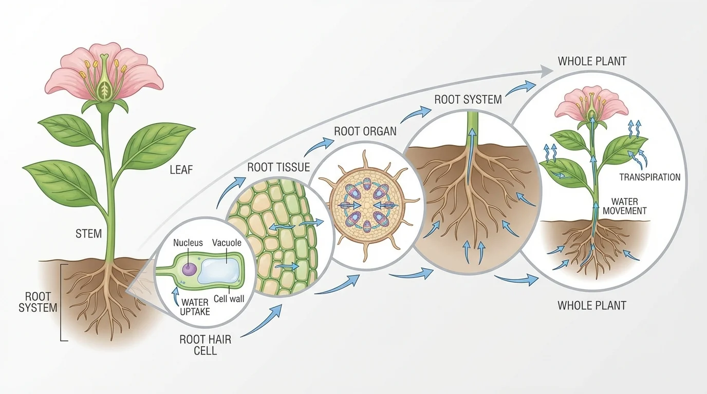
A model for a plant might show water moving up from root hair cells, through root tissues, into the root organ, then up the stem into leaves. Another part of the model could show how sugars produced in the leaves travel down to roots, illustrating interactions between these systems.
Other multicellular animals — from insects to whales — display similar hierarchies. Their specific tissues and organs differ, but the pattern of levels and interacting systems is shared. This universality is one reason biologists are confident that hierarchical organization is a fundamental principle of life.
When you are asked to "develop and use a model to illustrate the hierarchical organization of interacting systems," the goal is not just to copy a textbook diagram. Instead, you should design a representation that makes important relationships easier to understand.
To construct a strong model:
After building your model, you should also evaluate it critically:
For instance, the hierarchy ladder in [Figure 1] is excellent for understanding levels, but it does not show how different systems like digestive and circulatory interact. A second, network-style diagram connecting multiple organ systems might complement it.
Engineers use a similar approach when designing medical devices or artificial organs. They have to understand how their device will interact with tissues, organs, and systems across the hierarchy, and they build models — physical prototypes, computer simulations, and diagrams — to predict and improve performance before using them in real patients.
Before working with detailed models of body systems, it helps to recall that all organisms, from simple to complex, rely on cells as their basic structural and functional units. Even the most advanced organ system cannot function without coordinated activity at the cell level.
Ultimately, the power of modeling hierarchical organization lies in its ability to connect small-scale structures with large-scale functions: from the arrangement of cells to the survival of the organism. When you can move confidently up and down these levels in your explanations, you are thinking like a biologist.