Your body began as a single cell. From that one fertilized egg, repeated rounds of growth and division produced the enormous number of cells that now make up your tissues, organs, and body systems. That fact is remarkable on its own, but the underlying explanation is even more impressive: most of those cells contain the same genetic instructions, yet some become neurons, some become skin, some become muscle, and some become blood. The growth of a multicellular organism depends on two linked processes: cells must divide accurately, and then many of them must specialize.
In multicellular organisms, growth does not happen because individual cells simply become larger and larger forever. Instead, many cells grow to a certain point and then divide. This process, called mitosis, allows an organism to increase its number of cells while preserving the genetic information needed for life. The result is not just a larger mass of cells, but a highly organized living system in which different parts work together to meet the needs of the whole organism.
A fertilized egg, also called a zygote, forms when sperm and egg combine. This single cell contains a complete set of genetic information for a new organism. In humans, that includes two versions of each chromosome type, one inherited from each parent. As the zygote divides again and again, each parent cell passes on an identical set of DNA to both daughter cells. This continuity is essential. If cells in the same body had completely different instructions, coordinated growth and function would be impossible.
Early development begins with rapid cell division. At first, the embryo may increase cell number faster than overall size. Later, both cell division and cell enlargement contribute to growth. Over time, these cells begin to organize into layers, tissues, and structures. A multicellular organism is therefore not built all at once; it is assembled step by step through cellular reproduction and specialization.
Mitosis is the process in which the nucleus divides so that one parent cell produces two daughter cells with identical genetic material. Cytokinesis is the division of the cytoplasm, which usually follows mitosis and physically separates the two new cells.
Daughter cells are the two cells formed from one parent cell. In normal mitosis, each daughter cell receives the same number and kinds of chromosomes as the parent cell.
Although mitosis is often discussed as if it were only about growth, it is equally important for maintenance. Skin cells are constantly replaced. Cells lining the digestive tract are renewed. Cells near a wound divide to help repair tissue. The body is dynamic, not static. Even in a fully grown organism, many cells are still being produced, lost, and replaced.
To understand mitosis, you need to understand what is being copied and separated. DNA carries hereditary information. It contains the instructions for building proteins, regulating cell activities, and passing traits from one generation to the next. In eukaryotic cells, DNA is packaged into chromosomes inside the nucleus.
Most body cells in a multicellular organism have chromosomes in pairs. One member of each pair comes from one parent, and the other comes from the other parent. These paired versions may carry slightly different variants of genes, called alleles, but they are still corresponding chromosomes. Before a cell divides, it must copy its DNA so that each daughter cell can receive a full set.
After DNA replication, each chromosome consists of two identical copies joined together. These copies are called sister chromatids. During mitosis, the sister chromatids separate and move to opposite sides of the cell. This is how one copied set becomes two matching sets.
Genes are segments of DNA that contain instructions for making functional products, often proteins. Proteins influence cell structure and cell behavior, so preserving DNA accurately during cell division is critical for normal growth and function.
Accuracy matters because every cell depends on genetic instructions. If a liver cell is going to function as part of the liver, it must receive the full DNA set needed to make the proteins that support liver activity. If a skin cell is going to become part of the epidermis, it must also inherit complete DNA. Mitosis does not choose which genes a cell gets; it ensures that each daughter cell receives the same genome.
The life of a dividing cell follows an organized pattern called the cell cycle. This sequence, shown in [Figure 1], includes growth, DNA replication, preparation for division, and finally the division of the nucleus and cell. Cells do not jump randomly into mitosis. They pass through stages that help ensure the right size, enough resources, and a complete copy of DNA.
A large part of the cell cycle is interphase. During interphase, the cell grows, performs normal functions, and duplicates its DNA. By the end of this period, every chromosome has been copied, creating sister chromatids. Even though interphase is not part of mitosis itself, it is essential because mitosis cannot correctly distribute DNA that has not first been copied.
Mitosis is commonly described in stages. In prophase, chromosomes condense and become more visible, and the spindle begins to form. In metaphase, chromosomes line up near the middle of the cell. In anaphase, sister chromatids separate and move to opposite poles. In telophase, new nuclear envelopes form around the separated chromosome sets. Then cytokinesis divides the cytoplasm, producing two daughter cells.
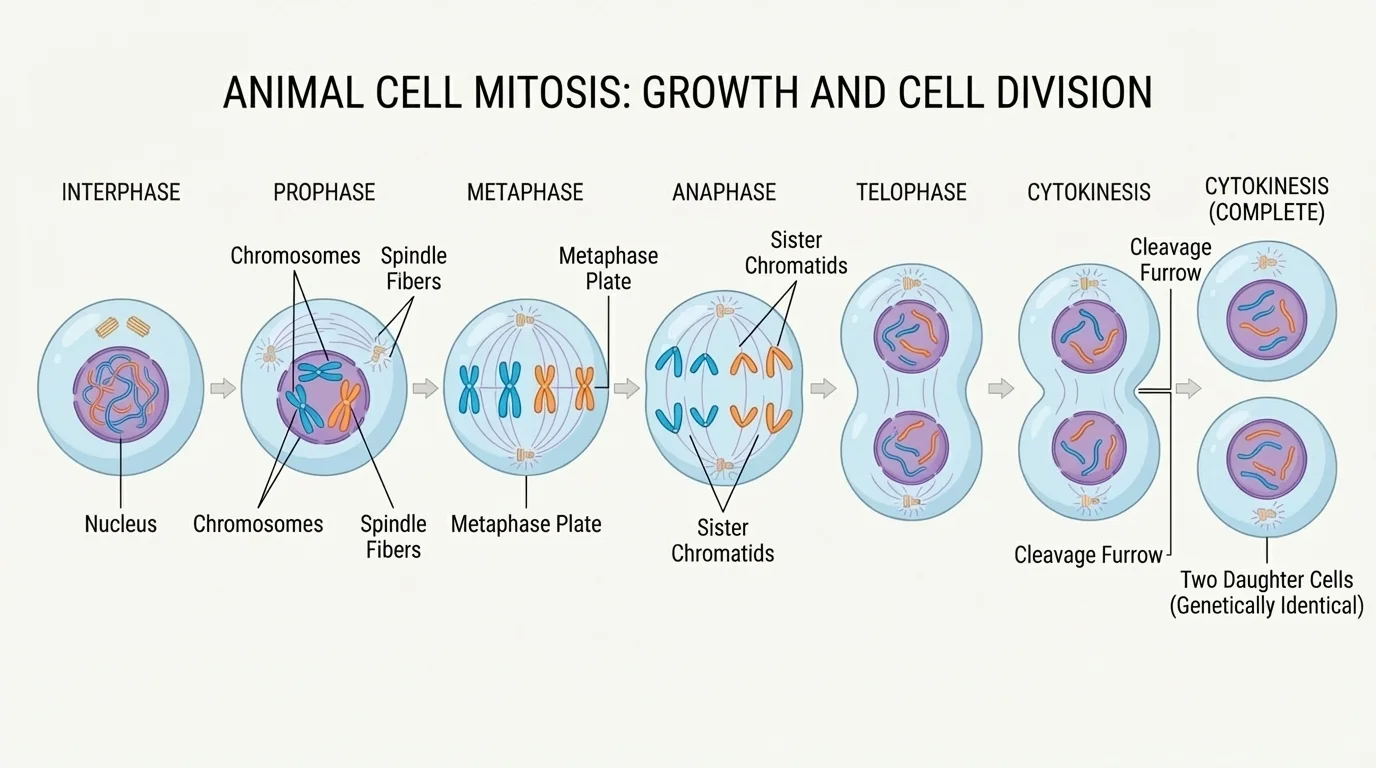
The spindle is especially important because it helps move chromosomes accurately. If chromosomes do not separate properly, daughter cells may receive too many or too few chromosomes. That can disrupt normal function or prevent survival. The precision of mitosis is one reason multicellular life can remain stable over countless rounds of cell division.
One useful way to think about mitosis is as an information-preserving process. The parent cell first copies its DNA, then sorts those copies into two equal groups. If a human body cell starts with a given number of chromosomes, each daughter cell should end with that same number. In symbolic form, if the parent cell has chromosome number \(n\), the daughter cells each also have chromosome number \(n\), because mitosis maintains chromosome number rather than reducing it. This is different from meiosis, which produces gametes and cuts chromosome number in half.
Later in development and tissue maintenance, the same pattern continues. A dividing skin cell, for example, still follows the stage sequence shown in [Figure 1]. The details may be microscopic, but the biological purpose is clear: accurate copying, accurate separation, and continued function.
Why mitosis produces genetically identical cells
Mitosis works because DNA is copied before division and because sister chromatids are separated evenly. If the replication step is accurate and the spindle separates chromatids correctly, each daughter cell receives the same genes in the same chromosome set as the parent cell. This allows growth and repair without changing the organism's basic genetic blueprint.
Cells do not divide nonstop. Chemical signals inside and outside the cell influence whether division should occur. Nutrients, growth factors, physical space, and signals from neighboring cells all help regulate the process. This regulation is vital for maintaining proper tissue size and structure.
During childhood and adolescence, growth depends heavily on mitosis. Bones lengthen because cells in growth regions divide. Muscle tissue can add cells in some contexts and increase cell size in others. Skin expands as the body grows because new cells are produced. The organism grows because cell number increases in a controlled way.
Mitosis also supports repair. When you scrape your skin, nearby cells divide to replace damaged ones. In the lining of the intestine, cells are frequently lost and replaced because the environment is harsh and cells wear out quickly. Blood-forming tissues in bone marrow continually produce new cells to replace those that age and die. These examples show that cell division is not only a developmental event but an ongoing necessity.
Real-world example: healing a cut
When skin is injured, repair depends on coordinated cell division and specialization.
Step 1: Cells near the wound detect signals released by damaged tissue.
Step 2: Some nearby cells enter the cell cycle and divide by mitosis.
Step 3: The new cells spread, cover the wound, and begin rebuilding the tissue structure.
Step 4: As healing continues, cells restore connections with surrounding tissue and normal function returns.
This is why a small cut can close over time without creating a completely new kind of tissue.
Not all tissues divide at the same rate. Some, like skin and intestinal lining, divide often. Others, like many neurons, divide very little or not at all once mature. This difference helps explain why some tissues heal faster than others. It also shows that multicellular organisms use cell division strategically, depending on tissue function.
Cell division alone cannot explain how a body becomes complex. It also depends on differentiation, the process by which unspecialized cells become specialized in structure and function. A neuron and a muscle cell usually contain the same DNA, yet they behave very differently because different genes are active in each cell type.
This difference in activity is called gene expression. A cell does not use all of its genes at once. Instead, it turns certain genes on and others off depending on its role, location, and signals from the environment. As a result, cells with the same genome can produce different proteins, develop different shapes, and carry out different jobs, as shown in [Figure 2].
Early in development, some cells remain relatively unspecialized and can still become multiple cell types. These cells are often described as stem cells. As development proceeds, many descendants become more restricted in what they can become. Eventually, specialized cells such as red blood cells, muscle fibers, and neurons perform highly specific functions.
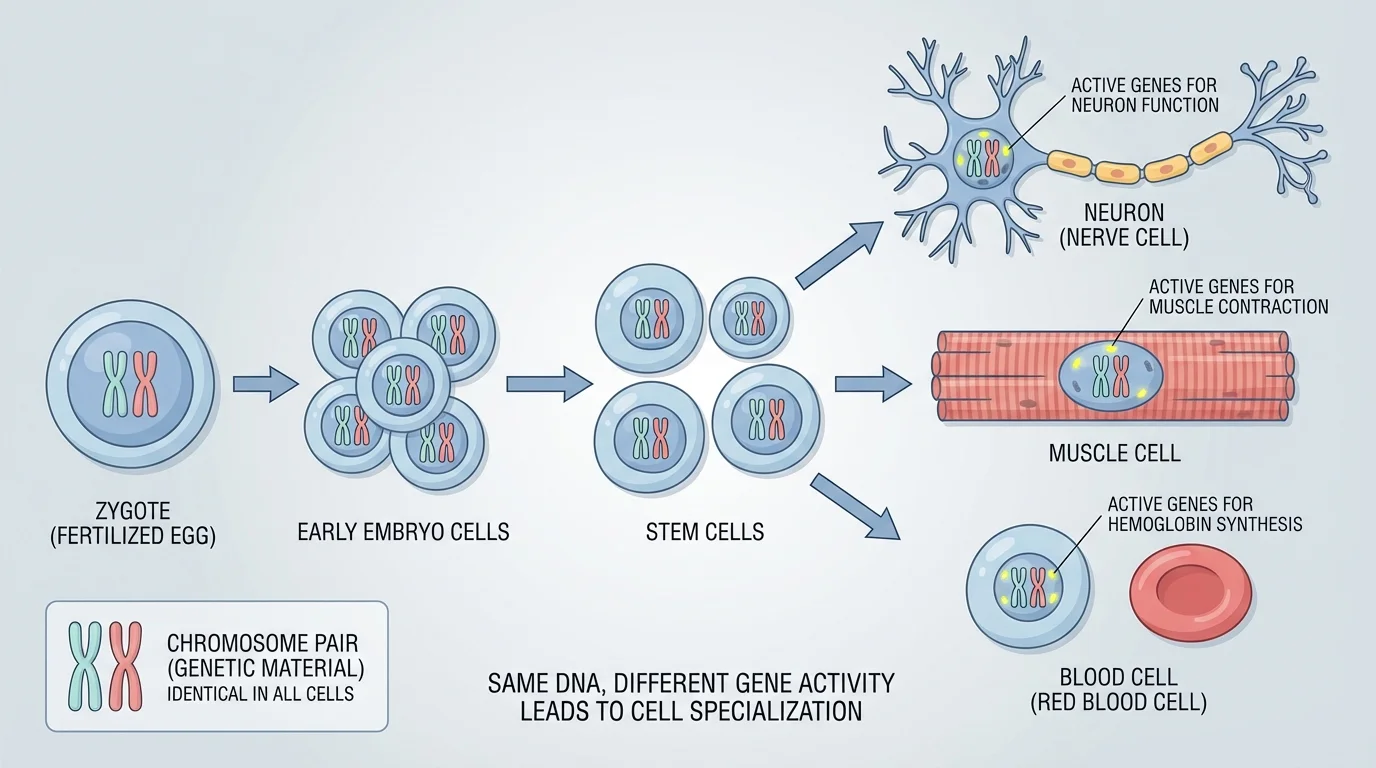
Differentiation is a major reason multicellular organisms can achieve complexity. A single type of cell could not efficiently digest food, carry oxygen, contract to move limbs, transmit electrical signals, and defend against pathogens all at once. Specialization divides labor across the organism, making life more efficient and more capable.
This specialization often involves visible structural differences. A neuron has long extensions that help transmit signals. A muscle cell contains abundant contractile proteins. Red blood cells are shaped to help transport oxygen efficiently. These cells look and behave differently because different sets of genes are expressed, even though they originated through repeated mitosis from earlier cells.
Some of the most powerful evidence for shared DNA in body cells comes from cloning and regenerative biology. In certain cases, the nucleus of a specialized cell still contains enough genetic information to direct the development of an entire organism if placed in the right environment.
The same developmental logic remains important throughout life. Tissue-specific stem cells in bone marrow, skin, and other organs continue producing new specialized cells when needed. In that sense, development never fully stops; it shifts from building the organism to maintaining it.
When comparing specialized cells, the pattern shown earlier in [Figure 2] remains essential: one lineage can branch into very different cell types without changing the fundamental fact that mitosis preserved the genome along the way.
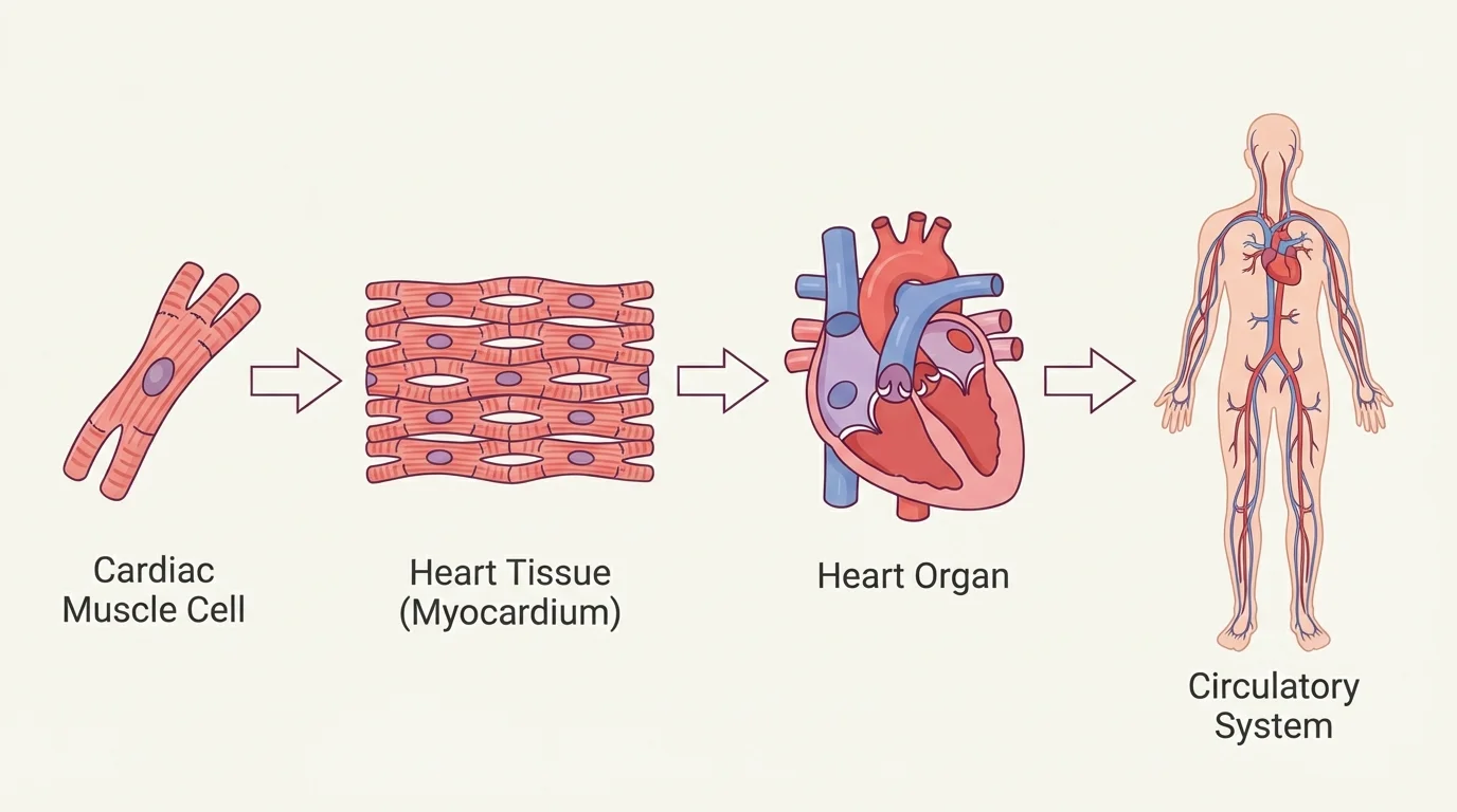
Multicellular life is organized in levels. Similar specialized cells form tissues. Different tissues combine to form organs, and organs work together in organ systems. This organization allows the body to solve large problems by coordinating many smaller parts.
Consider the heart, as shown in [Figure 3]. Cardiac muscle cells are specialized for contraction. Groups of these cells form cardiac muscle tissue. The heart also contains connective tissue, nervous tissue, and blood vessels. Together, these tissues form an organ that pumps blood. The heart then works within the circulatory system, alongside blood vessels and blood, to move oxygen, nutrients, hormones, and wastes throughout the body.
The same pattern appears across the body. Epithelial cells form protective linings. Muscle tissues produce movement. Nervous tissues transmit information. Connective tissues support and connect structures. Organs such as the lungs, kidneys, and stomach each contain multiple tissue types arranged for a specific function.
Organ systems are interdependent. The digestive system breaks food into absorbable molecules. The circulatory system transports those molecules. The respiratory system provides oxygen and removes carbon dioxide. The nervous and endocrine systems regulate timing and coordination. No organ acts entirely alone. The organism survives because systems work together.
| Level of organization | What it is | Example |
|---|---|---|
| Cell | Basic unit of life | Cardiac muscle cell |
| Tissue | Group of similar cells working together | Cardiac muscle tissue |
| Organ | Structure made of multiple tissues | Heart |
| Organ system | Group of organs coordinating major functions | Circulatory system |
| Organism | The whole living individual | Human |
Table 1. Levels of biological organization in a multicellular organism.
The hierarchy in [Figure 3] helps explain why cell division and differentiation matter beyond the microscopic scale. If mitosis failed, tissues could not be maintained. If differentiation failed, organs would lack the specialized parts needed to function. The health of the whole organism depends on both processes working together.
Because mitosis is so important, cells have checkpoint systems that help monitor DNA quality, chromosome attachment, and readiness to divide. These controls reduce errors. Still, mistakes can happen. DNA may be damaged by radiation, certain chemicals, replication errors, or other factors. If a damaged cell continues dividing, problems can spread through its descendants.
One serious outcome is cancer, a condition in which cells divide uncontrollably and ignore normal regulatory signals. Cancer cells may form tumors, invade nearby tissues, and sometimes spread to other parts of the body. In many cases, cancer develops after changes accumulate in genes that control the cell cycle, DNA repair, or cell death.
Why cancer is a disease of the cell cycle
Healthy tissues balance cell division, specialization, and cell death. Cancer disrupts that balance. Cells may continue through the cell cycle when they should stop, fail to repair DNA damage, or avoid signals that normally trigger cell death. The result is growth without proper control or contribution to the organism's needs.
Cancer also reveals why multicellular organisms require cooperation at the cellular level. In a healthy body, cells divide when needed and stop when appropriate. They function as part of a larger whole. Cancer cells act more like rule-breakers within that community, using resources without supporting the organism.
Not every cell-cycle error causes cancer, but all such errors remind us that growth is only beneficial when it is regulated. Biology is not simply about making more cells. It is about making the right cells, in the right place, at the right time, in the right numbers.
Modern medicine uses knowledge of mitosis and differentiation in many ways. Cancer treatments often target rapidly dividing cells. Bone marrow transplants rely on stem cells that can rebuild blood-forming tissues. Developmental biology helps scientists understand birth defects, tissue formation, and how organs emerge during embryonic growth.
Regenerative medicine explores how damaged tissues might be repaired more effectively. Researchers study how stem cells are guided into specific cell types and how growth signals can be controlled. This work has implications for treating burns, blood disorders, spinal cord injuries, and degenerative diseases.
These ideas also matter in agriculture and biotechnology. Plant growth depends on active regions of cell division called meristems. Tissue culture techniques can produce genetically similar plants from small samples, showing once again how a few cells can generate complex multicellular structures under the right conditions.
Real-world example: bone marrow and blood cell production
Bone marrow contains stem cells that help maintain the blood system.
Step 1: A blood-forming stem cell divides by mitosis, preserving the cell population.
Step 2: Some descendant cells begin differentiation.
Step 3: These cells develop into red blood cells, white blood cells, or platelets.
Step 4: The circulatory and immune systems continue functioning because old cells are replaced by new specialized ones.
This is a clear example of cell division and differentiation maintaining a complex organism throughout life.
Seen together, these processes explain one of the most important patterns in biology: a multicellular organism begins as one cell, expands through repeated mitosis, and becomes complex through differentiation and organization. Growth, maintenance, and coordinated function all depend on the faithful passing of genetic material and the controlled specialization of cells.