We all went through the process of embryo development. By the end of this lesson, you will describe the development of the embryo from the first cells to the blastocyst and understand complete details about the structures, development of the human embryo at different stages.
An embryo is the earliest stage in the development of a fertilized egg (the zygote) of a diploid, multicellular eukaryotic organism. It is the term used for any animal or plant, from the first cell division until birth, or hatching, or germination in plants.
In humans, it is called an embryo until about eight weeks after fertilization, and from then until birth it is called a fetus. The development of the embryo is called embryogenesis, and the study of embryos is called embryology.
Generally in organisms reproducing sexually a zygote develops into an embryo. A zygote is a single cell that is as a result of fertilization of the egg cell of the female by the sperm cell of the male. A zygote has got half of the DNA from both the parents. In some protists, animals, and plants, the zygote begins division through mitosis leading to the production of an organism that is multicellular. This results in an embryo.
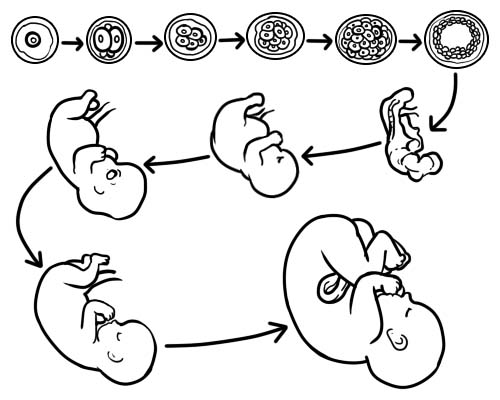
The development of an embryo from the zygote takes place through certain stages of organogenesis, blastula, and gastrula.
The first stage is the blastula stage. It is characterized by a fluid-filled cavity referred to as the blastocoel. Around this is a sphere of cells which are referred to as the blastomeres. In mammals having a placenta, fertilization of the ovum takes place in the fallopian tube via which it moves into the uterus. The word fetus is used to refer to a more advanced embryo’s stage of development that runs until hatching or otherwise birth. This occurs from week eleven of gestation in humans. In animals, however, the development of eggs takes place outside the body of the mother, are called embryos throughout their development. For example, chick embryos are not referred to as chick fetuses regardless of their development stage.
During the second stage, gastrulation, the blastula cells undergo cell division’s coordinated processes. Apart from the process of cell division, they also undergo other processes such as invasion as well as migration that leads to the formation of either two or three layers of tissue. When two layers are formed it is referred to as diploblastic and when three layers are formed it is referred to as triploblastic. In organisms that are triploblastic, the germ layers are the endoderm, mesoderm, and ectoderm. The germ layer’s position and arrangement, are highly species-specific but this depends on the type of the produced embryo. A special embryonic cell population exists in vertebrates which are referred to as the Neural crest. It is now the fourth germ layer after being proposed. It is believed to have been of importance in the head structure's evolution.
During the third stage, organogenesis, cellular and molecular interactions between the germ layers, combined together with the developmental potential of the cells, responding competence, lead to further differentiation of cell types that are organ-specific. For example, in the process of neurogenesis, an ectoderm subpopulation of cells differentiates to become peripheral nerves, spinal cord, and the brain. Modern biology is seeking to understand the molecular basis for all organogenesis types including chondrogenesis (formation of cartilage), osteogenesis (formation of bones), myogenesis (formation of muscle) and angiogenesis (new blood vessels formation from pre-existing ones).