Your skin looks smooth, a leaf looks flat, and pond water may look like plain greenish liquid. But those everyday things are hiding a busy world that your eyes cannot detect. A single drop of pond water can contain living organisms, and a thin slice of onion can reveal box-like structures that prove plants are made of tiny units of life. One of the biggest ideas in science is that what you can observe depends on the scale at which you look.
In science, scale means the size level you are observing. You might look at something at the level of a whole organism, such as a tree, a dog, or your own body. You might also look at a much smaller level, such as tissues, cells, or even molecules. Some features are easy to notice at one scale but completely invisible at another.
For example, from far away, a forest may look like one green blanket. As you move closer, you can distinguish separate trees. Closer still, you can see leaves. With magnification, you can see cells in the leaf. At an even smaller level, you could study molecules such as \(\textrm{H}_2\textrm{O}\), proteins, and \(\textrm{CO}_2\) involved in life processes. Each level reveals new information, but it also hides other information. A whole forest pattern cannot be seen when you are staring at one leaf cell.
Scale is the size level at which something is observed. A cell is the basic unit of structure and function in living things. A microscope is a tool that makes very small objects appear larger so they can be studied.
This idea matters because scientists must choose the right scale to answer the right question. If you want to know whether a tree is healthy, you may inspect leaves, bark, and growth. If you want to know why a leaf is losing water, you may need to look at cells and tiny openings in the leaf. If you want to know how the leaf makes sugar, you may need to study molecules such as \(\textrm{CO}_2\) and \(\textrm{H}_2\textrm{O}\) inside chloroplasts.
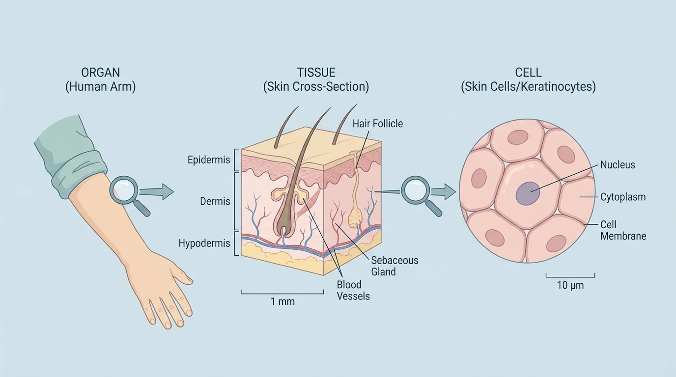
One of the central ideas in biology is that all living things are made of cells. This may seem obvious for tiny organisms, but it is also true for very large organisms. Your body contains many trillions of cells. A whale, an oak tree, and a mushroom are also made of cells. The smooth skin on your arm may not look cell-based, but it is actually built from many small living units.
[Figure 1] This idea is part of cell theory, which states that living things are made of one or more cells, the cell is the basic unit of life, and new cells come from existing cells. Students in middle school often focus especially on the first part: every living thing is made of cells.
Some organisms consist of just one cell. Others consist of many cells with many different types. Even though cells are tiny, they are not just empty bubbles. Each cell contains structures that help it carry out life functions such as obtaining energy, growing, responding, and reproducing.
When scientists investigate a slice of onion, cells from the inside of the cheek, or microorganisms in pond water, they gather evidence for this idea directly. The evidence is visual: instead of seeing a solid mass, they observe separate units with boundaries. In plants, these units often look like tiny boxes because of their cell walls. In animal cells, the edges are usually softer and less box-like.
Notice how the evidence changes with scale. With the unaided eye, an onion slice looks smooth and thin. Under magnification, repeated cell shapes appear. The phenomenon "this onion is made of cells" is not observable at the larger scale of everyday vision, but it becomes observable at the cellular scale.
Some single-celled organisms can do everything needed for life within one cell: move, get energy, respond to the environment, and reproduce. That means one cell can be a complete organism.
This is why scientists say that observations depend on tools and scale, not just on whether something is real. Cells are present whether you can see them or not.
Your eyes are powerful, but they have limits. A grain of sand is visible to you, but most cells are far too small to be seen clearly without help. To study life at smaller scales, scientists use tools. The choice of tool depends on the question, and the same sample reveals different details as magnification increases.
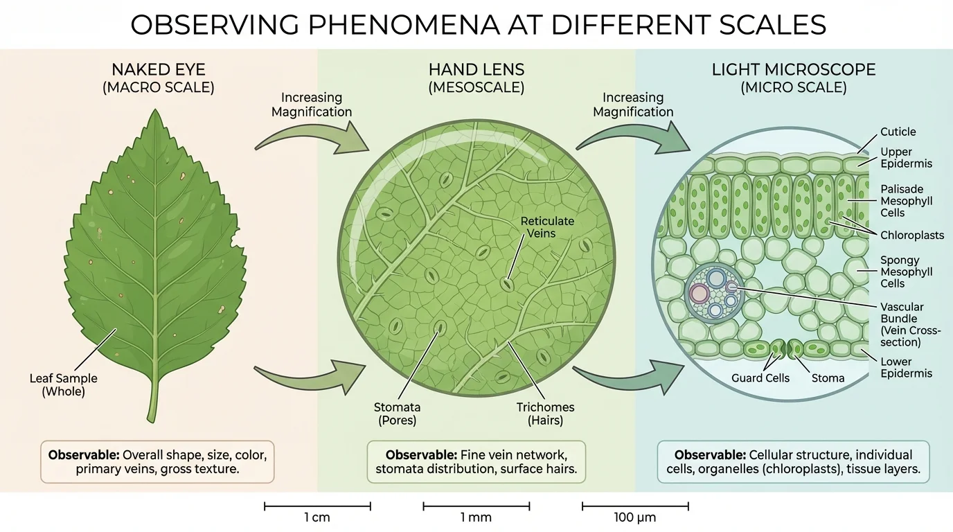
[Figure 2] A hand lens can enlarge small objects such as insect wings, leaf veins, or the texture of fabric. It helps you notice features that are too small for normal viewing but still not tiny enough to require a microscope.
A light microscope uses light and lenses to magnify objects to a much greater extent. With a classroom microscope, students can often see plant cells, cheek cells, and many microorganisms. They may also notice parts such as the cell membrane, nucleus in some cells, or chloroplasts in plant cells.
Magnification increases the apparent size of an object, while resolution is the ability to see two nearby points as separate. A blurry enlargement is not enough; scientists need enough detail to distinguish structures. A microscope works best when the sample is thin enough for light to pass through.
This helps explain why some life processes are hard to understand from everyday observation alone. For example, a cut on your skin heals over time. At the organism scale, you see the wound close. At the cellular scale, cells are dividing and replacing damaged cells. At the molecular scale, proteins are helping rebuild tissue. Each scale provides part of the story.
An organism made of one cell is called unicellular. Examples include many bacteria and some protists. A unicellular organism carries out all life processes in one cell. It may move with tiny structures, absorb nutrients, remove waste, and reproduce, all without specialized body parts.
An organism made of many cells is called multicellular. Humans, fish, trees, birds, and most fungi are multicellular. In these organisms, different cells often have different jobs. That allows the organism to become larger and more complex.
Why being multicellular changes what can be observed
At the whole-organism scale, a multicellular organism may show behaviors such as running, growing, or reacting to light. At the cellular scale, those same actions depend on many cells working together. The larger-scale behavior is visible to the eye, but the cooperation among cells is not visible without magnification and deeper study.
A single-celled organism may be invisible in pond water, yet under a microscope it becomes a complete living thing. A multicellular organism like a maple tree is easy to see from far away, but the cells that make up its bark, leaves, and roots are hidden. This is a strong example of how one scale can reveal an organism while another reveals its building blocks.
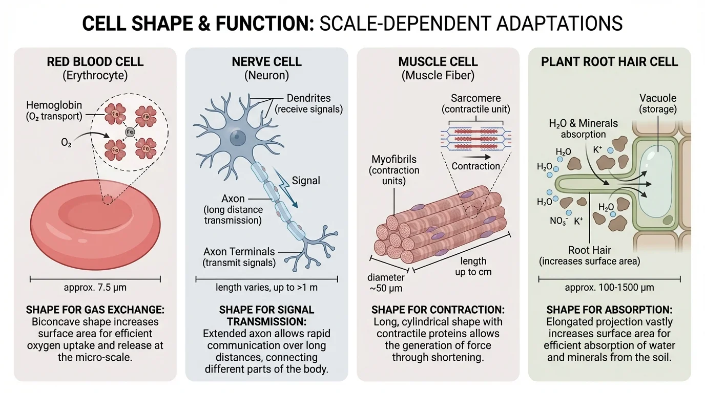
In multicellular organisms, cells are not all identical. They become specialized, meaning they develop structures that help them perform specific tasks. A cell's shape often matches its function. A long nerve cell helps carry signals over distance, while a red blood cell is shaped to move easily through blood vessels and transport gases.
[Figure 3] Plant cells can also specialize. Root hair cells have long extensions that increase surface area for absorbing water and minerals. Leaf cells contain many chloroplasts to capture light energy. Guard cells open and close tiny pores to regulate gas exchange and water loss.
At the scale of the whole organism, you may observe that muscles contract, roots absorb water, and leaves make food. At the cellular scale, different cell types are carrying out the work. The phenomenon "the body has parts with different functions" is visible on a large scale, but the reason behind it often becomes clearer only when you study specialized cells.
Cells are organized into tissues, tissues into organs, and organs into organ systems. This organization means that patterns repeat at different scales. Many similar muscle cells form muscle tissue. Different tissues combine to form a heart. The heart works with blood vessels to form part of the circulatory system.
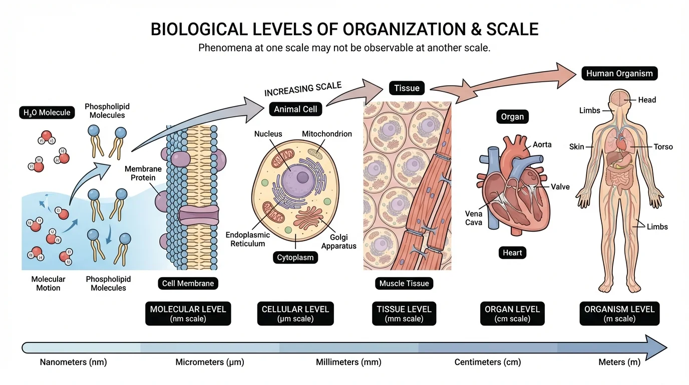
Life can be studied at many levels of organization, and this idea can be presented as a connected hierarchy from molecules to organisms. Each level has structures and processes that are easier to observe there than anywhere else.
[Figure 4] At the molecule level, scientists study substances such as \(\textrm{H}_2\textrm{O}\), \(\textrm{O}_2\), \(\textrm{CO}_2\), lipids, proteins, and DNA. Molecules are much too small to see with classroom microscopes, but they are essential for understanding how cells work. Photosynthesis, for example, involves molecules moving and reacting inside cells.
At the cellular level, you can observe boundaries, internal structures, movement in some organisms, and differences among cell types. This is the level where students often gather direct evidence that living things are made of cells.
At the tissue and organ levels, patterns become larger and easier to recognize. You can identify leaf veins, skin layers, heart chambers, or stem structures. These are groups of cells working together.
At the organism level, you can observe visible traits such as size, shape, behavior, growth, and response to the environment. You can watch a plant bend toward light or an animal run away from danger, but you cannot directly see the molecular reactions causing those responses.
This means no single scale gives the entire picture. If you only observe a whole leaf, you might understand its shape and color but not how chloroplasts capture light. If you only observe chloroplasts, you might miss how the leaf is arranged on the plant to collect sunlight. Science often involves moving back and forth between scales.
| Level of observation | What can often be observed | What may remain hidden |
|---|---|---|
| Organism | Behavior, overall shape, growth | Individual cells and molecules |
| Organ | Parts such as leaf, heart, root | Most cell details |
| Tissue | Groups of similar cells | Molecular activity |
| Cell | Cell shape, boundaries, some organelles | Most molecules |
| Molecule | Chemical composition and reactions | Whole-organism patterns |
Table 1. Different biological scales reveal different kinds of information while hiding other details.
Earlier, [Figure 1] showed how tissue is built from many cells. That same idea helps explain why injuries, growth, and disease can look simple from the outside but involve countless cell-level events inside the body.
Scientists do not just state that living things are made of cells; they collect evidence. In a classroom investigation, students may prepare a thin onion peel, add a stain, and examine it under a light microscope. They may also look at cheek cells or a drop of pond water.
Example investigation: observing onion cells
Step 1: Prepare a very thin layer of onion tissue on a slide.
Step 2: Add a drop of stain to make structures easier to see.
Step 3: Place a coverslip carefully over the sample.
Step 4: Observe under low power, then higher power on the microscope.
Step 5: Record evidence such as repeated box-like compartments, clear boundaries, and arrangement of many separate units.
The repeated compartments are evidence that the onion tissue is made of cells, not one solid sheet.
A good investigation depends on clear evidence. If students see many repeating units with boundaries, that supports the idea that the sample is cellular. If they switch from low magnification to higher magnification, they can compare what becomes visible at each scale. Using the right scale improves the quality of the evidence.
Cheek cells give different evidence. They do not look box-like like onion cells, but they still appear as separate living units. Pond water adds another variation: students may find single-celled organisms that are entire organisms by themselves. In one investigation, students can gather evidence for both multicellular and unicellular life.
All organisms need energy, respond to their environment, grow, and reproduce. Cells are the level at which these life functions are carried out.
As seen earlier in [Figure 2], changing the tool changes what can be observed. A hand lens may help with surface texture, but only a microscope reveals most cells clearly enough to use as evidence.
Doctors and medical scientists constantly work across scales. A physician may observe symptoms at the organism level, such as fever, coughing, or swelling. A laboratory scientist may then examine cells, tissues, or molecules to discover the cause. An infection can look like one illness at the body scale, but under a microscope it may involve bacteria, damaged cells, and immune cells interacting.
Cancer is another example. At the large scale, a doctor may detect a lump or unusual tissue growth. At the cellular scale, cancer involves cells dividing in an uncontrolled way. At the molecular scale, changes in DNA can affect how those cells behave. Understanding the disease requires linking all three scales.
Plant scientists also use scale. Farmers may notice that crops are yellowing or wilting. Looking more closely at root cells, leaf tissues, or nutrient molecules helps explain why. A problem visible in the field may begin at a much smaller scale.
Microscopes changed science so much that many ideas about disease and life had to be revised after scientists could finally observe cells and microorganisms directly.
Biotechnology depends on this scale shift too. Scientists may grow cells in laboratories, edit genes, or study proteins, then apply what they learn to improve medicine, agriculture, and environmental protection. What is invisible to the eye can still have major effects on everyday life.
One misunderstanding is that if something cannot be seen directly, it is less important or less real. In biology, some of the most important processes happen at scales too small for the eye to detect. Cells divide, membranes control what enters and leaves, and molecules store and transfer information.
Another misunderstanding is that zooming in always gives a better understanding. Zooming in gives different information, not always more useful information. If you want to know whether a bird can migrate, you study the organism and its behavior. If you want to know how its muscles release energy, you must study cells and molecules.
A third misunderstanding is that all cells look the same. As [Figure 3] demonstrates, cells vary in shape and structure because different forms support different jobs. In multicellular organisms, this variation is one reason complex life is possible.
Finally, students sometimes think there is a sharp break between levels of organization. In reality, levels are connected. Molecules build cell parts. Cells form tissues. Tissues form organs. Organs work together in organisms. Observing one level helps explain the next.
"To understand life, scientists often have to zoom in and zoom out."
The most powerful scientific explanations connect scales. A plant bends toward light because of behavior seen at the organism level, growth in tissues, changes in cells, and chemical signals at the molecular level. None of these observations alone is the complete story.