Your body is carrying out billions of events right now without asking your permission: oxygen is moving into blood, glucose is being broken down in cells, hormones are signaling organs, and neurons are firing in patterns that let you read this sentence. No scientist can watch all of that directly at every scale at once. That is why biology depends on models. Models make the invisible understandable.
In science, a model is not just a tiny copy of something. A model is any representation that helps us explain, visualize, test, or predict how a system works. In biology, models help us study systems made of many interacting parts, from molecules inside a cell to organ systems inside an entire organism.
When scientists model living systems, they often focus on three kinds of flow: energy flow, matter flow, and information flow. Energy powers processes such as muscle contraction and active transport. Matter includes substances such as oxygen, water, nutrients, and wastes moving through the body. Information includes electrical impulses, chemical signals, and feedback messages that coordinate responses.
System means a set of interacting parts that work together. A biological system may be a cell, an organ, an organ system, or an entire organism.
Model means a representation of an object, process, or system used to describe, explain, or predict what happens.
Simulation means using a model, often mathematical or computer-based, to imitate how a system changes over time.
Biology is especially rich in systems because multicellular organisms are built from levels of organization that depend on one another. Understanding those levels is essential for understanding why models are so useful.
Scientists use models because real biological systems are often too small, too large, too fast, too slow, or too complex to study directly all at once. A heart can be dissected, but that does not reveal every pressure change during exercise. A cell can be viewed under a microscope, but there are too many molecular interactions to track by eye. A model simplifies reality so that key patterns stand out.
There are several major types of models. Physical models are tangible objects, such as a plastic heart, a skeleton, or a 3D-printed lung. Mathematical models use quantities and equations to describe relationships. Computer models combine data, rules, and calculations to simulate changes over time.
No model is perfect. Every model leaves something out. That is not a flaw by itself; it is a choice. A good model includes the details needed to answer a question and leaves out details that would make the model harder to use without improving understanding.
Modern medicine depends heavily on modeling. Doctors use computer models to estimate blood flow, predict the spread of infectious disease, and even plan surgeries using patient-specific scans.
Because models simplify, scientists must always ask what a model captures well and what it misses. That habit becomes especially important when we study the organization of living bodies.
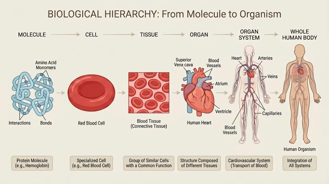
A multicellular organism is a hierarchy of interacting parts, as [Figure 1] shows. The body is not just a pile of cells. Molecules form structures inside cells, cells form tissues, tissues form organs, and organs work together in organ systems. Each level has specific functions, but each also depends on levels above and below it.
A protein molecule such as hemoglobin helps red blood cells carry oxygen. Red blood cells are part of blood tissue. Blood travels through organs such as the heart and blood vessels. Those organs make up the circulatory system, which interacts with the respiratory and digestive systems to support the whole organism. A change at one level can affect all the others.
This hierarchical organization explains why biology often needs multiple models rather than one giant all-purpose model. A model of a protein explains binding and shape. A model of a cell explains transport and metabolism. A model of an organ system explains coordinated function. Each reveals part of the same living system.
Consider muscle movement. At the molecular level, actin and myosin proteins interact. At the cellular level, muscle cells shorten. At the tissue level, muscle fibers pull together. At the organ level, a skeletal muscle contracts. At the organ-system level, muscles, bones, and nerves coordinate movement. If you study only one level, you miss important causes and effects.
The same logic applies across the body. The stomach does not function alone; it depends on nerves, hormones, blood supply, smooth muscle, and epithelial tissue. A model that only shows the organ's shape is useful for anatomy, but not enough for explaining digestion.
Hierarchy and function
Biological organization is hierarchical because structures at smaller scales combine to create functions at larger scales. The key idea is interaction: cells do not simply exist next to one another; they communicate, exchange materials, and coordinate activities to produce tissue and organ function.
As we move through these levels, what flows through them also matters. Organisms stay alive because substances, energy, and signals keep moving.
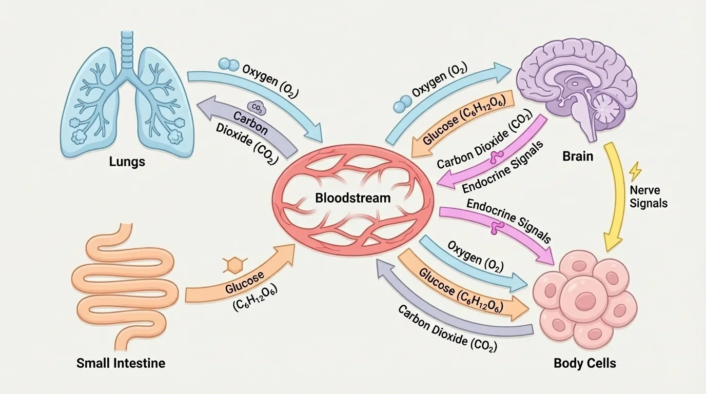
Within a living organism, survival depends on continuous movement of materials and messages, and [Figure 2] helps make that connection visible across systems. Matter flows when molecules such as glucose, carbon dioxide, water, and ions move from one place to another. Energy flows when chemical energy stored in food is transformed into ATP and then used for cellular work. Information flows when receptors detect change and send signals through the nervous or endocrine systems.
Take cellular respiration as an example. Cells use cellular respiration to release energy from glucose. A simplified chemical equation is
\[\textrm{C}_6\textrm{H}_{12}\textrm{O}_6 + 6\textrm{O}_2 \rightarrow 6\textrm{CO}_2 + 6\textrm{H}_2\textrm{O} + \textrm{energy}\]
Here, matter is rearranged: glucose and oxygen become carbon dioxide and water. Energy is transferred from chemical bonds into forms the cell can use. This process links the digestive system, respiratory system, circulatory system, and cells throughout the body.
For example, the digestive system breaks food into absorbable molecules. The small intestine moves nutrients into the blood. The respiratory system brings oxygen into the lungs, where it diffuses into blood. The circulatory system delivers these materials to cells. Cells release carbon dioxide, which blood carries back to the lungs for exhalation.
Information flow is just as important. If blood glucose rises after a meal, the pancreas detects that change and releases insulin. Cells respond by taking in more glucose. This is a signaling process, not just a transport process. A useful model must often include both matter movement and information control.
Energy transformations can also be modeled numerically. If one molecule of glucose stores a certain amount of chemical energy, cells do not use all of it with perfect efficiency. Some energy becomes heat. That is why your body temperature rises during vigorous exercise. Energy flow in biology always involves transformation, not magic creation.
Numerical example: estimating oxygen use during exercise
A student's tissues use about \(0.30 \textrm{ L}\) of oxygen per minute at rest and \(2.40 \textrm{ L}\) per minute during intense exercise.
Step 1: Compare the two rates.
The factor increase is \(\dfrac{2.40}{0.30} = 8\).
Step 2: Interpret the result.
The body needs oxygen at a rate that is 8 times greater during intense exercise than at rest.
This simple mathematical model helps explain why breathing rate and heart rate increase together.
The body does not treat these flows separately. During exercise, as we saw conceptually in [Figure 2], respiratory, circulatory, muscular, and nervous systems all interact at once.
A physical model is often the easiest kind to understand first because you can see and touch it. In a classroom or lab, physical models may show the chambers of the heart, the branching of bronchi in the lungs, or the arrangement of the digestive tract.
Physical models are especially useful for showing shape, size, spatial arrangement, and connections among structures. A 3D model of the nephron, for example, can help students trace how filtrate moves through kidney structures. A torso model can show how the diaphragm, lungs, heart, liver, and stomach fit together in limited space.
However, physical models usually have limits. They often show structure better than change. A plastic heart may clearly show valves and vessels, but it does not naturally show pressure waves, electrical conduction, or changing blood oxygen levels. To study those, scientists turn to mathematical or computer models.
Structure and function are closely linked in biology. If a model shows only shape, ask what function that shape makes possible. If a model shows only function, ask what structures are responsible.
Even so, physical models remain valuable in medicine. Surgeons now use 3D-printed models made from imaging data to plan difficult procedures. These models can represent an individual patient's anatomy, which is far more useful than a generic diagram in certain cases.
Mathematical models reveal patterns that are hard to notice by intuition alone, and [Figure 3] highlights one of the most important biological examples: the relationship between surface area and volume. This relationship helps explain why cells remain small and why multicellular organisms need specialized transport systems.
For a cube-shaped cell with side length \(s\), the surface area is \(6s^2\) and the volume is \(s^3\). The surface area to volume ratio is therefore
\[\frac{6s^2}{s^3} = \frac{6}{s}\]
As \(s\) gets larger, the ratio gets smaller. That means larger cells have less surface area available per unit volume for exchanging materials with the environment.
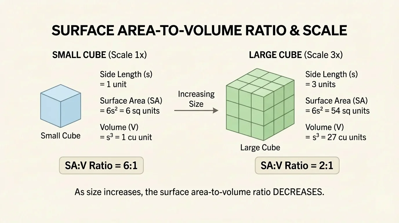
Worked example: surface area to volume ratio
Compare two cube-shaped cells, one with side length \(1 \textrm{ unit}\) and one with side length \(3 \textrm{ units}\).
Step 1: Find the smaller cell's surface area and volume.
Surface area: \(6(1^2) = 6\). Volume: \(1^3 = 1\). Ratio: \(\dfrac{6}{1} = 6\).
Step 2: Find the larger cell's surface area and volume.
Surface area: \(6(3^2) = 54\). Volume: \(3^3 = 27\). Ratio: \(\dfrac{54}{27} = 2\).
Step 3: Interpret the comparison.
The larger cell has more total surface area, but much less surface area relative to its volume. This makes exchange less efficient.
This helps explain why most cells are small and why larger organisms rely on circulatory systems instead of simple diffusion alone.
Another mathematical model can describe rate. If blood flow is estimated by volume per time, then a heart pumping \(70 \textrm{ mL}\) per beat at \(72\) beats per minute has a cardiac output of
\[70 \times 72 = 5{,}040 \textrm{ mL/min}\]
That is about \(5.04 \textrm{ L/min}\). If exercise raises heart rate and stroke volume, output rises, helping deliver more oxygen and nutrients. This model does not capture everything about circulation, but it gives a useful estimate.
| Model type | What it represents well | What it may miss |
|---|---|---|
| Physical | Shape, position, structure | Time-based changes, invisible flows |
| Mathematical | Rates, ratios, quantitative relationships | Anatomical detail, full biological complexity |
| Computer | Dynamic interactions, feedback, many variables | Depends strongly on assumptions and data quality |
Table 1. Comparison of major model types used to study biological systems.
The surface area to volume relationship in [Figure 3] also helps explain organ design. Lungs and intestines increase surface area through branching and folding, which improves exchange without requiring a huge increase in overall body size.
A computer model can track many variables at once and update them over time. That makes it ideal for studying feedback, disease spread, organ interactions, and multiscale biological systems. Computer simulations are especially useful when direct experimentation is too slow, too dangerous, or ethically impossible.
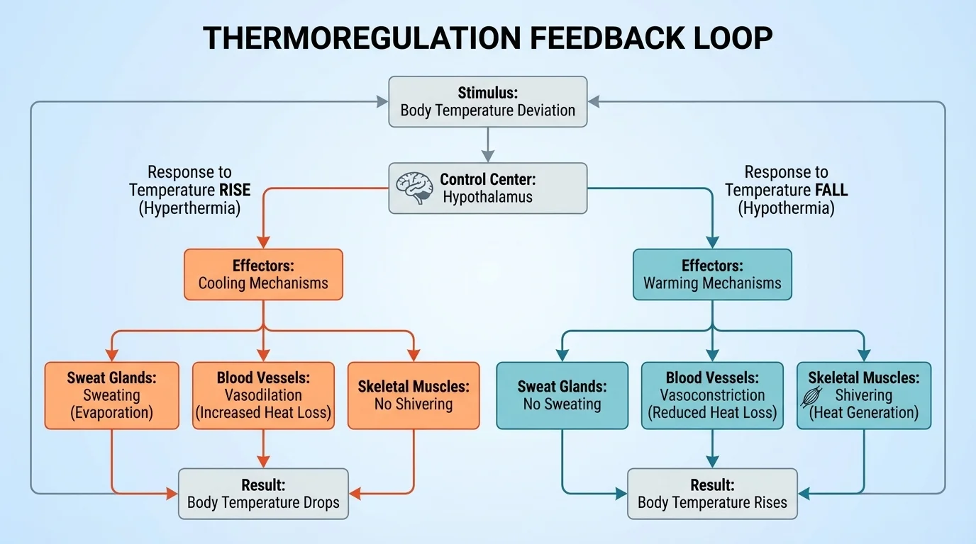
One major idea in biology is homeostasis, the maintenance of relatively stable internal conditions. Computer simulations of homeostasis often include sensors, control centers, and effectors, as [Figure 4] illustrates for body temperature regulation. If temperature rises, the system responds with sweating and blood vessel dilation. If temperature falls, shivering and blood vessel constriction help conserve or generate heat.
These models are powerful because they can show time. A physical model of the body can show where sweat glands are located, but a computer simulation can show how temperature changes minute by minute as external temperature, activity level, and blood flow change.
Computer models are also used in epidemiology to predict how diseases spread through populations, in neuroscience to simulate networks of neurons, and in pharmacology to estimate how a drug concentration changes in blood and tissues. These are not separate from organismal biology; they connect directly to how systems interact within and between organisms.
Case study: simulating blood glucose control
A simplified computer model of blood glucose regulation may include glucose intake from food, insulin release from the pancreas, glucose uptake by cells, and storage in the liver.
Step 1: A meal raises blood glucose.
The model increases the glucose variable after carbohydrate absorption from the small intestine.
Step 2: The pancreas responds.
If glucose rises above a set level, the model increases insulin output.
Step 3: Target tissues respond.
Cells take in more glucose, and the liver stores more as glycogen.
Step 4: The level falls toward normal.
The simulation shows negative feedback because the response reduces the original change.
This kind of model helps scientists study diabetes and test treatment strategies.
Later, the same feedback pattern seen in [Figure 4] appears in many systems, including blood pressure regulation, calcium balance, and water balance.
When you sprint, several systems interact immediately. Muscles need more ATP. The respiratory system increases air intake. The circulatory system speeds delivery of oxygen and nutrients. The nervous system coordinates movement and adjusts heart rate. The endocrine system releases hormones such as adrenaline. Modeling this event requires more than one organ diagram because the body behaves as a coordinated network.
This is why scientists often build linked models. One part may represent the lungs, another the heart, another the blood, and another the cells using oxygen. Inputs and outputs connect them. For example, more carbon dioxide in blood can trigger changes in breathing rate, which changes oxygen supply, which affects cellular respiration. Information flow controls matter flow, and both affect energy availability.
Medical monitoring devices also rely on model-based thinking. A pulse oximeter estimates blood oxygen saturation using light absorption. It does not directly count every oxygen molecule. Instead, it uses a model connecting light signals to hemoglobin behavior. Every reading on the screen is based on a simplified representation of the real system.
Interactions across organ systems
Functions in multicellular organisms emerge from system interactions, not isolated parts. Digestion supplies nutrients, respiration supplies oxygen, circulation transports materials, excretion removes wastes, and nervous and endocrine systems coordinate responses. A useful model often needs to represent these links, not just the parts themselves.
Exercise, stress, dehydration, illness, and injury all reveal these interconnections. A problem in one system can spread effects through many others. For example, reduced kidney function can alter water balance, blood pressure, ion concentration, and heart workload.
A biological process can look very different depending on the scale being studied. At the molecular level, insulin is a signaling molecule binding to receptors. At the cellular level, membranes move glucose transporters. At the tissue level, liver and muscle tissue change how they store or release glucose. At the organism level, blood glucose concentration rises or falls. The same event can require several models at different scales.
Scientists choose scale based on the question. If the question is why a mutation changes a protein's shape, a molecular model is best. If the question is why a patient feels fatigued, a whole-body systems model may be more useful. The challenge is knowing when to zoom in and when to zoom out.
This is also why one model should not be mistaken for reality itself. A diagram of the circulatory system may show major vessels clearly, but it cannot capture every capillary and every chemical exchange. A computer model of the immune system may simulate millions of interactions, but it still relies on assumptions. Scientific understanding improves when models are compared, tested, and revised.
Some of the most advanced biological models are now built from real patient data, including MRI scans, blood chemistry, and genetic information. These personalized models are helping develop more targeted treatments.
Across scales, the key pattern remains the same: living systems are organized, interactive, and constantly exchanging energy, matter, and information.
To evaluate a model, scientists ask several questions. What parts of the system are included? What has been simplified? What evidence supports the model? Does it explain observations? Can it predict what will happen under new conditions?
A strong model is not necessarily the most detailed one. It is the one that is most useful for a particular purpose. A classroom lung model made of bottles and balloons may successfully show pressure changes during breathing. It does not need to include every alveolus to explain inhalation and exhalation.
Models are revised when new evidence appears. If a simulation predicts that a medicine should lower blood pressure but clinical results differ, scientists examine the assumptions, add missing variables, or change the equations. In this way, modeling is not separate from experimentation; it works alongside it.
Biology becomes much clearer when you think in terms of systems. Molecules, cells, tissues, organs, and organ systems are not isolated chapters in a textbook. They are levels of one interacting structure. Models help us trace how a change at one level affects the others and how energy, matter, and information keep the organism functioning.