Your body is made of trillions of cells, yet most of them are far too small to see with your eyes alone. That sounds almost impossible: how can something invisible be so important? Scientists solve this problem by using microscopes, diagrams, and models. These tools help us study tiny systems and understand a big scientific idea: the way something is built helps explain what it does.
Many natural systems are too small, too large, too fast, or too complex to understand all at once. A cell is a great example. It is microscopic, but it is also a busy system with many parts working together. As shown in [Figure 1], scientists use a model to represent the cell and make its structures easier to observe and explain. A model might be a drawing, a 3D object, a computer simulation, or even a labeled chart.
Models are helpful because they can make the invisible visible. They can enlarge tiny structures, simplify complicated details, and show relationships among parts. For example, a textbook picture of a cell is not an exact photograph. It is a model that helps you notice important structures such as the membrane, nucleus, and mitochondria.
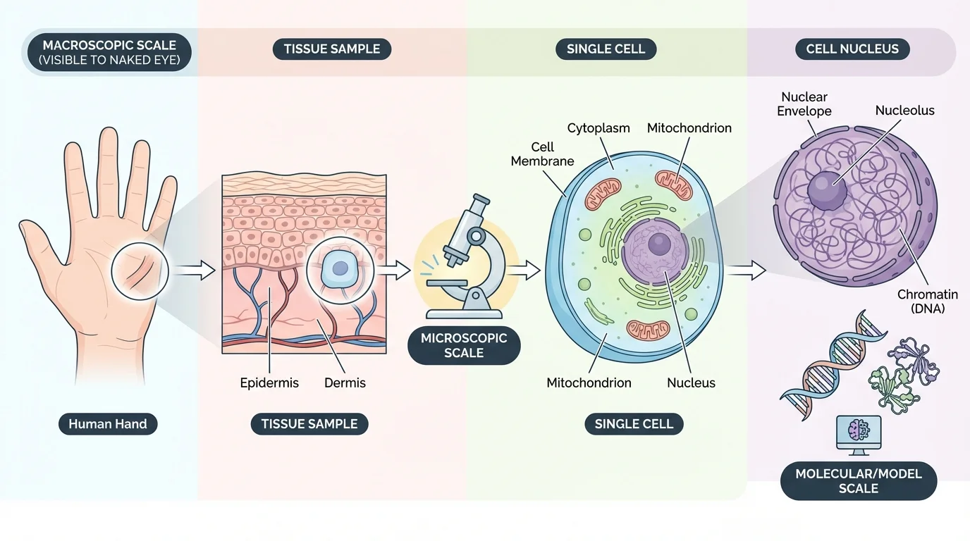
Scientists also use different kinds of microscopes. A light microscope can show whole cells and some larger parts. More powerful microscopes can reveal even smaller details. But even microscope images need interpretation. A scientist often compares an image with a model to figure out which structures are present and what they might be doing.
Structure and function means that the shape, material, arrangement, and size of a part help determine what that part can do. In living systems, this idea helps scientists explain why cell parts look different and perform different jobs.
This same idea applies beyond cells. Bird beaks, leaf shapes, bones, and blood vessels all have forms that fit their jobs. When scientists analyze a natural system, they ask questions such as: What are the parts made of? How are the parts arranged? How do the parts interact? Those questions help reveal how the system functions.
All living things are made of one or more cells. Some organisms, such as many bacteria, are made of just one cell. Others, such as humans, oak trees, and mushrooms, are made of many cells. A cell is the basic unit of life because it can carry out the processes needed for living, including getting energy, growing, responding to the environment, and reproducing.
Even though cells are small, they are not simple blobs. Each cell has organized parts that help it survive. In multicellular organisms, cells can also specialize. Muscle cells are shaped to contract. Nerve cells are shaped to send signals over long distances. Leaf cells are arranged to capture light. Their structures are connected to their functions.
Living things need energy, matter, and information. Cells take in materials, use energy to power reactions, and follow genetic instructions stored in DNA. These basic ideas help explain why cells need different internal parts.
A single cell can be viewed as a complete system. Like a city, it has boundaries, transportation routes, power sources, storage spaces, instructions, and waste removal. This comparison is not perfect, but it helps show that a cell works because its parts are connected rather than isolated.
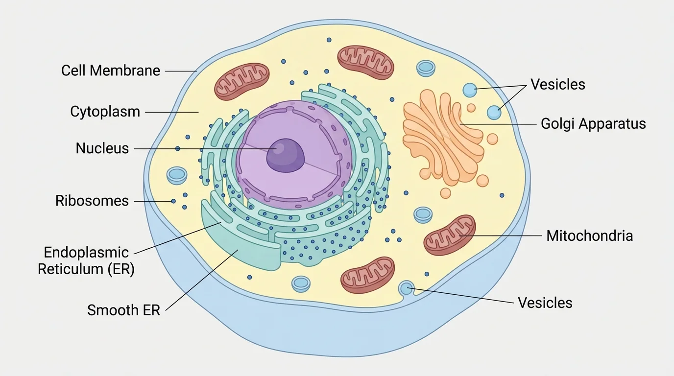
As illustrated in [Figure 2], when scientists study a cell, they do not just list its parts. They analyze how each organelle contributes to the work of the whole cell. Organelles are small structures inside cells that perform specific functions. Their relationships matter just as much as the parts themselves.
For example, the nucleus stores instructions, ribosomes build proteins, the endoplasmic reticulum helps process and move materials, and the Golgi apparatus packages them. Mitochondria release usable energy from food. The cell membrane controls what enters and leaves. If one major part fails, the whole system is affected.
This is why scientists talk about cells as systems. A system is a group of interacting parts that work together. In a cell, no organelle does everything. Function depends on cooperation. The shape and placement of each part help the cell work efficiently.
Thinking in systems also helps explain disease. If a membrane is damaged, materials may leak in or out. If mitochondria do not work well, the cell may not have enough energy. If DNA is altered, the cell may make the wrong proteins. Studying the system helps scientists identify what went wrong.
The cell membrane forms the outer boundary of the cell. It is thin and flexible, but it is also highly selective. This means it controls which substances can enter and which can leave. That control is essential because the cell must take in nutrients, release waste, and maintain stable internal conditions.
The membrane is made mostly of lipids and proteins. Its structure allows some small molecules, such as \(O_2\) and \(CO_2\), to pass through more easily than large molecules. Proteins in the membrane act like gates, channels, or helpers that move specific substances across. The membrane's structure fits its job: a protective boundary that still allows communication and exchange.
Inside the membrane is the cytoplasm, a jelly-like region where organelles are suspended. Many chemical reactions happen there. The cytoplasm helps support the organelles and allows materials to move within the cell. It is not empty space. It is an active environment where cell life happens.
Real-world connection: Why membrane structure matters
Step 1: Consider a sports drink entering the digestive system.
Water and dissolved substances move toward body cells, but they cannot simply rush into every cell all at once.
Step 2: The cell membrane controls entry.
Membrane proteins help certain particles move across while blocking others. This keeps the cell from swelling, shrinking, or losing important materials.
Step 3: Link structure to function.
Because the membrane is thin, flexible, and packed with specialized proteins, it can protect the cell and regulate transport at the same time.
The cell survives because its boundary is not just a wall. It is a working control surface.
The cell membrane also helps cells communicate. Some membrane proteins receive signals from other cells. In a multicellular organism, this communication allows cells to coordinate activities. For example, cells in the immune system recognize signs of infection partly through structures at their membranes.
The nucleus is often called the control center of the cell because it contains most of the cell's DNA. DNA carries the instructions for building proteins, and proteins influence traits and cell activities. The nucleus does not control the cell by magic. It does so by storing and managing information.
This idea connects to inheritance. Organisms pass genetic information from parents to offspring. Those instructions help determine traits such as eye color, leaf shape, or certain inherited conditions. Cells use the information in DNA to make molecules that support life. When DNA changes, cell function can change too.
DNA links structure, function, and inheritance. DNA is a molecule with a specific structure that allows it to store coded information. That information guides the building of proteins. Proteins help form cell structures and carry out chemical jobs. Because DNA can be copied and passed on, traits can be inherited from one generation to the next.
The nucleus is surrounded by a membrane with openings called pores. These openings allow certain materials to move in and out. That structure matters. The DNA must be protected, but instructions also need to be shared with other parts of the cell. The nucleus therefore balances protection with communication.
Cells that are very active in making proteins may have a nucleus that appears especially busy because genetic information is constantly being used. As we saw earlier in [Figure 2], the nucleus is not working alone. It is part of a larger system that includes ribosomes and transport structures.
One of the most important jobs of a cell is making proteins. Proteins help build structures, speed up reactions, send signals, and transport substances. As shown in [Figure 3], the cell does not do this in one place. It uses a connected production system that works like a cellular assembly line.
Ribosomes are the structures where proteins are built. Some float freely in the cytoplasm, while others attach to the rough endoplasmic reticulum. The rough endoplasmic reticulum, often shortened to rough ER, has ribosomes on its surface and helps process and transport newly made proteins.
The smooth ER does not have ribosomes. It helps make lipids and performs other tasks such as detoxifying certain chemicals. This difference in surface appearance matches a difference in function. Rough ER is specialized for protein processing, while smooth ER handles other jobs.
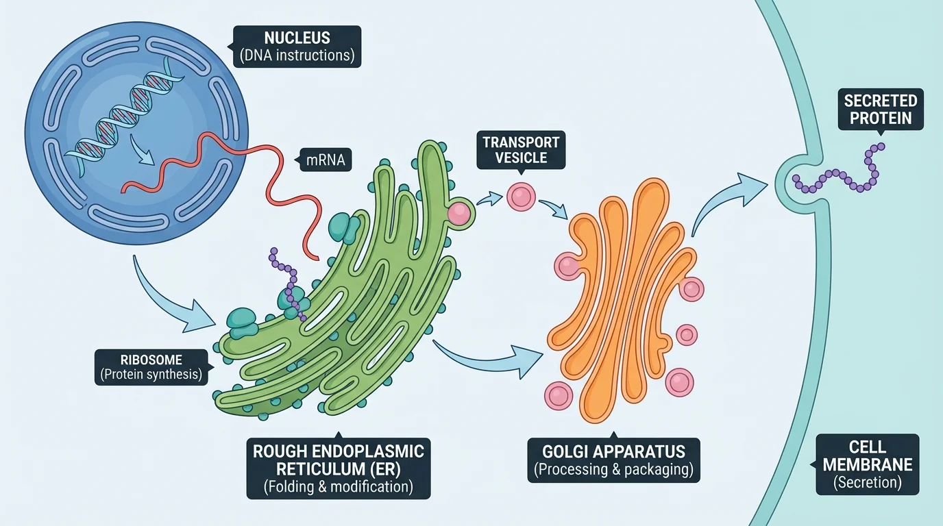
The Golgi apparatus receives molecules from the ER, modifies them, sorts them, and packages them into vesicles. Vesicles are small membrane-bound sacs that move materials around the cell. This arrangement is efficient because each structure handles part of the process. The parts form a pathway rather than acting independently.
A skin cell, for example, needs proteins that help protect the body. The DNA instructions are used to build proteins at ribosomes, those proteins move through the ER, the Golgi apparatus packages them, and vesicles deliver them to where they are needed. Later, when scientists study secretion or transport problems, they often refer back to pathways like the one in [Figure 3].
As shown in [Figure 4], cells need energy to carry out life processes. The mitochondrion helps release usable energy from food molecules. Its inner membrane is highly folded, and those folds increase surface area. More surface area allows more of the reactions involved in energy release to take place. Here, shape directly supports function.
Plant cells and some other organisms also contain chloroplasts. Chloroplasts capture light energy and use it to help make sugar. They contain chlorophyll, the pigment that gives plants their green color. Inside chloroplasts are stacks of membranes that help with the reactions of photosynthesis.

A simplified description of photosynthesis is: \[6CO_2 + 6H_2O + \textrm{light energy} \rightarrow C_6H_{12}O_6 + 6O_2\]. In words, carbon dioxide and water are used to make glucose and oxygen using light energy. For example, if a plant cell uses \(6\) molecules of \(CO_2\) and \(6\) molecules of \(H_2O\), it can produce \(1\) molecule of \(C_6H_{12}O_6\) and \(6\) molecules of \(O_2\) in this balanced model.
Mitochondria and chloroplasts both have membranes arranged in ways that maximize important reactions. Their internal designs are different, but in both cases the folded or stacked surfaces help the organelle do more work efficiently.
Plants use chloroplasts to make sugars, and both plant and animal cells use mitochondria to release energy from those sugars. That connection links two major cell processes. The products of one process can become the inputs of another.
A vacuole is a storage structure. Vacuoles can hold water, nutrients, or waste. Plant cells often have one large central vacuole that stores water and helps support the cell. When the vacuole is full, it pushes outward, helping the plant stay firm.
Plant cells also have a cell wall, a rigid outer layer outside the membrane. The cell wall gives support and protection. Its stiffness helps plants stand upright even though they do not have bones. This is another example of structure and function: a rigid material supports the needs of the organism.
These plant cell features fit the life of a plant. Plants cannot move to search for food, so they use chloroplasts to make it. They need support to reach sunlight, so cell walls and full vacuoles help keep stems and leaves upright. Their parts make sense when you think about the jobs plants must do.
Plant and animal cells share many basic structures because both are eukaryotic cells. They have nuclei, membranes, cytoplasm, ribosomes, mitochondria, ER, and Golgi apparatus. But they also have important differences, and [Figure 5] makes those differences easier to compare side by side.
The table below compares major features of plant and animal cells.
| Cell Structure | Plant Cell | Animal Cell | Main Function |
|---|---|---|---|
| Cell membrane | Yes | Yes | Controls movement of materials |
| Nucleus | Yes | Yes | Stores DNA and instructions |
| Cytoplasm | Yes | Yes | Site of many reactions |
| Mitochondria | Yes | Yes | Release usable energy |
| Chloroplasts | Usually yes | No | Capture light energy for photosynthesis |
| Cell wall | Yes | No | Support and protection |
| Large central vacuole | Usually yes | No | Storage and support |
Table 1. Comparison of major structures in plant and animal cells and their functions.
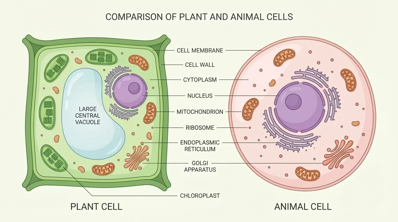
Animal cells often have more flexible shapes because they do not have a rigid cell wall. That flexibility is useful in tissues such as muscle or blood. Plant cells, in contrast, often look more box-like because the cell wall gives them a more fixed shape. Later, when studying tissues and whole organisms, the comparison in [Figure 5] helps explain why plant bodies and animal bodies are built differently.
Cells do not exist only as isolated units. In multicellular organisms, similar cells work together to form tissues, tissues form organs, and organs form organ systems. The structures inside each cell help the larger body function. For instance, muscle tissue depends on cells that use lots of energy, so those cells often contain many mitochondria.
Leaf tissue contains many chloroplast-rich cells because leaves are major sites of photosynthesis. Root cells may have structures that help absorb water and minerals. In each case, cell structure supports tissue function, and tissue function supports the organism.
Case study: Why a leaf is shaped the way it is
Step 1: Look at the job of the leaf.
A leaf needs to capture light, exchange gases, and move water and sugars.
Step 2: Look at the cells inside it.
Many leaf cells contain chloroplasts. Their arrangement allows light to reach photosynthetic tissues.
Step 3: Connect small structures to larger function.
Because chloroplasts, membranes, and transport tissues are arranged effectively, the entire leaf can function as a food-making organ.
This is how scientists analyze complex systems: they connect the structures of parts to the functions of systems.
The same thinking works in human biology. Red blood cells have a shape that helps them move through blood vessels and carry oxygen. Nerve cells are long so they can transmit signals across long distances. Structure and function connect at every level of life.
Models are powerful, but they are not perfect copies of reality. A cell model might leave out tiny details, show colors that are not natural, or make some parts larger than they really are. Even so, models are useful because they highlight key relationships and make patterns easier to notice.
Scientists often compare several models at once. They might use microscope images, computer renderings, and hand-drawn diagrams. Each model emphasizes something different. One may show shape well, another may show movement, and another may simplify the system enough for students to understand the main idea.
This approach is especially helpful in medicine. Cancer researchers study how changes in cell structure affect function. Virologists examine how viruses enter cells through membrane interactions. Geneticists analyze how DNA changes affect proteins and traits. In each case, understanding the parts and their relationships helps explain the behavior of the whole system.
"The way a thing is built tells us a great deal about what it can do."
— A core idea of biology
When scientists ask why a system works, they often begin by mapping its parts. Then they test how changing one part affects the rest. That is true for cells, ecosystems, machines, and even weather systems. Complex systems become understandable when we visualize them and analyze the connections within them.
Understanding cell structure has practical value. Doctors study damaged cells to diagnose disease. Farmers use knowledge of plant cells to improve crop growth. Biotechnologists grow cells in laboratories to make medicines, including some vaccines and proteins used in treatment.
For example, if a scientist wants to know why a certain plant survives drought better than another, they may examine the plant's cells. Do the cells store water effectively? Are the membranes maintaining balance well? Are the chloroplasts functioning under heat stress? Looking at the microscopic structures helps answer a larger real-world question.
Engineers also borrow ideas from biology. Membranes inspire filtration technology. Branching structures in organisms can inspire designs for transportation and cooling systems. By studying how natural structures work, scientists and engineers learn how to solve human problems.