Your body is not a collection of separate parts working alone. Every breath, every heartbeat, every movement depends on communication and exchange across many levels of organization. A single muscle contraction in your leg depends on molecules, cells, tissues, nerves, blood vessels, and organs working together with astonishing precision. Biology becomes much clearer when you stop looking at parts in isolation and start seeing the relationships between them.
Scientists often use models to understand these relationships. A model can be a drawing, diagram, physical object, computer simulation, or even a mathematical description. In biology, models are especially useful because living systems are complex. A good model does not include every detail. Instead, it focuses on the components and interactions that matter for explaining a function, such as how oxygen gets to cells, how food becomes usable energy, or how the body keeps internal conditions stable.
A biological system is a group of interacting parts that work together to carry out a function. The respiratory system brings in oxygen. The circulatory system transports materials. The nervous system sends rapid signals. None of these systems is completely independent. Their functions overlap and support one another.
When scientists build models, they use evidence from observations and experiments. Evidence can include microscope images, medical scans, measurements of heart rate, oxygen concentration, blood glucose levels, and many other types of data. A model based on evidence is stronger than a guess because it is supported by what has actually been observed.
Model in biology means a simplified representation used to explain or predict how a structure, process, or system works. Evidence-based means the model is supported by data such as observations, experiments, measurements, or images.
One reason models are powerful is that they reveal patterns. For example, many body systems follow the same logic: materials move, signals are exchanged, and feedback helps maintain stability. Once you recognize these patterns, complex systems become easier to understand.
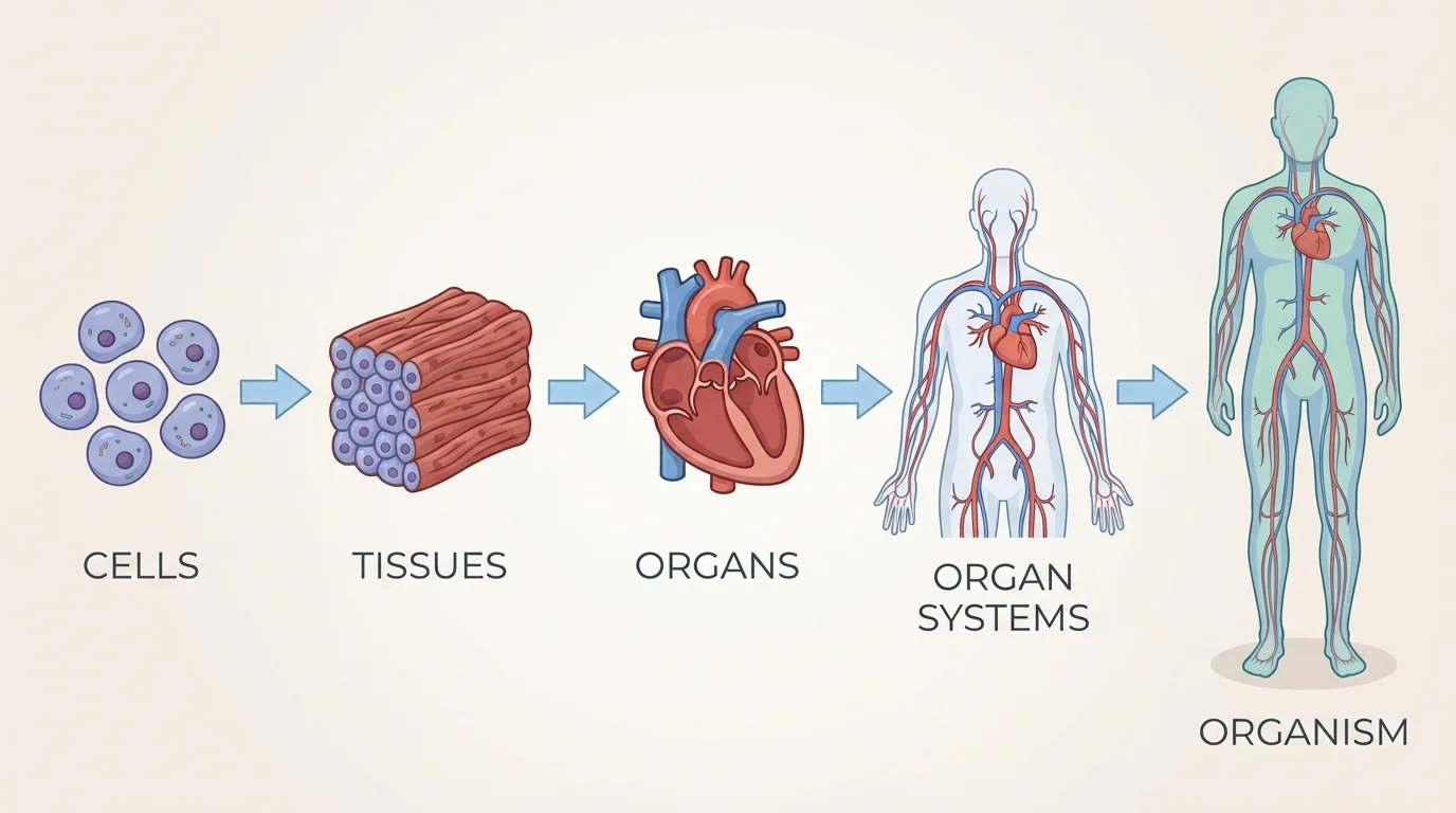
In multicellular organisms, structure is arranged in nested levels, as [Figure 1] shows. Small components become parts of larger components, and each level contributes to specific functions. This hierarchy helps scientists organize what they know and identify where a particular process occurs.
The basic living unit is the cell. Similar cells working together form a tissue. Different tissues combine to make an organ. Organs that cooperate form an organ system. All organ systems together make the organism. For example, muscle cells form muscle tissue, muscle tissue helps make the heart, and the heart is part of the circulatory system.
This hierarchy matters because function depends on relationships between levels. A heart is not just cardiac muscle. It also contains connective tissue, nerve tissue, blood vessels, and specialized cells that generate electrical signals. Its ability to pump blood comes from how these parts interact, not from one part alone.
Consider the stomach. Its epithelial tissue secretes digestive chemicals, smooth muscle tissue churns food, nerve tissue helps control contractions, and blood vessels supply oxygen and carry absorbed substances away. A model of the stomach that ignored these relationships would miss how the organ really works.
The same idea applies across the whole body. Your organism-level traits, such as endurance, growth, and temperature control, emerge from coordinated interactions across many smaller levels. Later, when we examine gas exchange and energy use, the hierarchical organization in [Figure 1] remains essential for understanding why body systems depend on one another.
A scientific model is not just an illustration made to look neat. It is a tool for explaining relationships. If a model shows arrows between the lungs and blood, those arrows should represent observed movement of gases. If it shows nerve signals affecting muscles, that relationship should be supported by experiments or recordings of electrical activity.
Evidence for biological models comes from many sources. Light and electron microscopy reveal cell structures. Imaging methods such as X-rays, MRI, ultrasound, and CT scans show organs and tissues in living bodies. Chemical tests measure concentrations of molecules like \(\textrm{O}_2\), \(\textrm{CO}_2\), and glucose. Pulse oximeters estimate blood oxygen saturation. These observations allow scientists to create models that connect structure with function.
Useful models are selective. A model should include the parts needed to answer a question. If the question is how oxygen reaches muscle cells during exercise, the model should include lungs, alveoli, blood vessels, the heart, red blood cells, and body tissues. It does not need to include every bone in the skeleton.
All models have limits. A textbook diagram may make organs look separate and tidy, even though real tissues are crowded, folded, and dynamic. A flowchart may show the direction of a process but not the speed. A computer simulation may predict trends but still simplify chemical details. Knowing a model's strengths and limits is part of scientific thinking.
One of the clearest examples of interacting systems is the movement of oxygen from the air to body cells, a pathway that [Figure 2] illustrates through linked components of the respiratory and circulatory systems. This is not just about breathing. It is about how structures at several levels make gas exchange and transport possible.
Air enters through the nose or mouth, travels through the trachea, and reaches the lungs. Inside the lungs are tiny air sacs called alveoli. Their walls are extremely thin and are surrounded by equally thin capillaries. This structure creates a short distance for diffusion, allowing oxygen to move from air in the alveoli into the blood, while carbon dioxide moves from blood into the alveoli to be exhaled.
The blood then carries oxygen to the heart, which pumps it through arteries to tissues throughout the body. At body cells, oxygen leaves the blood and enters cells, where it is used in reactions that release energy from food molecules. Carbon dioxide produced by cells moves into the blood and is carried back to the lungs.
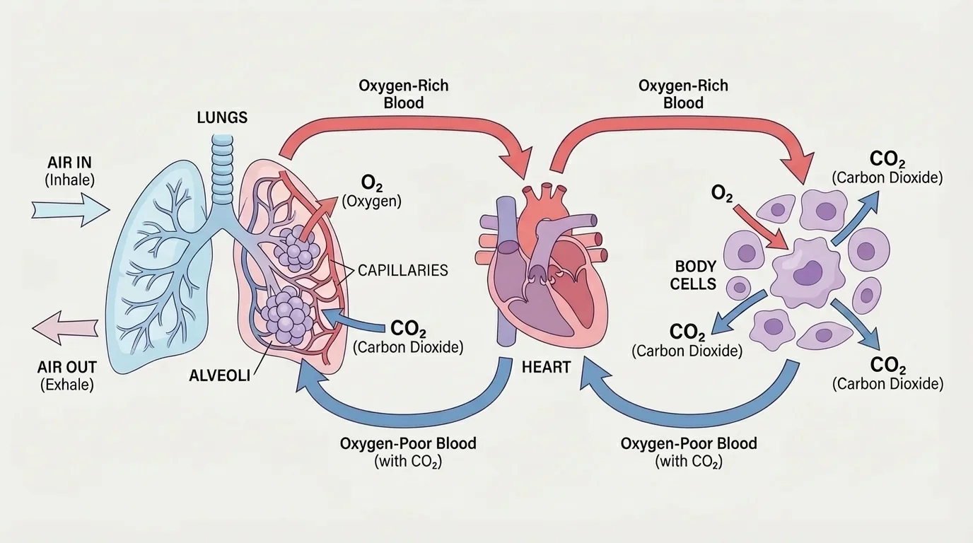
This system works because the components match their functions. Alveoli provide large surface area. Capillaries bring blood close to air spaces. Red blood cells contain hemoglobin, which binds oxygen. The heart generates the pressure needed to move blood. If you modeled only the lungs and ignored blood flow, the explanation would be incomplete.
Here is a simple relationship often used in physiology: the amount of oxygen delivered to tissues per minute depends on cardiac output and oxygen content of the blood. In simplified form, oxygen delivery follows the pattern \(\textrm{delivery} = \textrm{blood flow} \times \textrm{oxygen content}\). For a numeric example, if blood flow is \(5 \textrm{ L/min}\) and oxygen content is \(200 \textrm{ mL/L}\), then oxygen delivery is \(5 \times 200 = 1{,}000 \textrm{ mL/min}\). The exact biology is more detailed, but this relation helps show why both breathing and circulation matter.
Real-world example: sprinting up stairs
When a person runs up several flights of stairs, both breathing rate and heart rate increase.
Step 1: Muscles need more energy, so their cells use oxygen faster and produce more carbon dioxide.
Step 2: The respiratory system increases ventilation so more air reaches the alveoli.
Step 3: The circulatory system increases blood flow so oxygen and glucose reach muscles more quickly.
Step 4: Carbon dioxide and heat are removed more rapidly, helping maintain internal balance.
This example shows why no single organ can explain exercise response by itself.
The relationships in [Figure 2] also help explain disease. In asthma, narrowed airways reduce airflow. In pneumonia, fluid in the lungs interferes with gas exchange. In heart failure, blood is not pumped efficiently enough to deliver materials. Different components fail, but the effect spreads through the entire system.
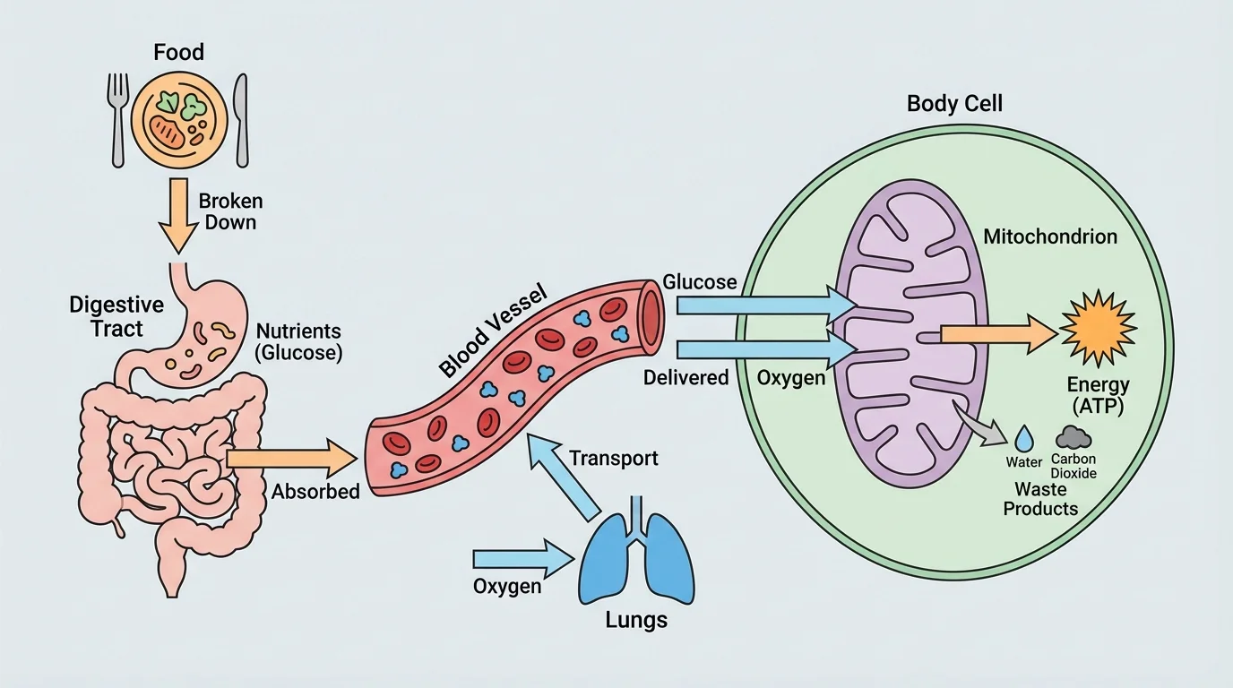
Another strong system model connects the digestive system to circulation and then to energy release in cells, as [Figure 3] shows. Food does not help you simply because it is in your stomach. Its molecules must be broken down, absorbed, transported, and used by cells.
In digestion, large food molecules are mechanically and chemically broken into smaller molecules. Carbohydrates are broken into sugars such as glucose. Proteins are broken into amino acids. Fats are broken into fatty acids and glycerol. Much of this absorption occurs in the small intestine, whose folded structure and tiny villi increase surface area for uptake.
Once glucose enters the bloodstream, the circulatory system transports it to cells throughout the body. Oxygen delivered by the respiratory and circulatory systems also enters those cells. In the mitochondria, cells use glucose and oxygen in cellular respiration to release usable energy. A simplified chemical equation is \(\textrm{C}_6\textrm{H}_{12}\textrm{O}_6 + 6\textrm{O}_2 \rightarrow 6\textrm{CO}_2 + 6\textrm{H}_2\textrm{O} + \textrm{energy}\).
This model explains why digestion alone is not enough. If the blood cannot carry nutrients, cells cannot use them. If oxygen supply is limited, cells cannot fully carry out aerobic respiration. If cells are damaged and cannot use the molecules delivered, energy production still falls.
A simple quantitative relationship helps here too: if one glucose molecule reacts with \(6\) oxygen molecules, then \(3\) glucose molecules would require \(18\) oxygen molecules because \(3 \times 6 = 18\). Students do not need every biochemical step to grasp the systems idea. The key point is that molecules entering one system become inputs for another.
The small intestine is several meters long and covered with folds, villi, and microvilli, creating enormous surface area for absorption. That structural design is one reason the digestive system can move huge numbers of molecules into the bloodstream efficiently.
The pathway in [Figure 3] becomes especially important in diabetes. When blood glucose regulation is disrupted, cells may not receive or use glucose properly even when food has been eaten. That problem begins with regulation, but its effects reach circulation, energy use, and organ function across the body.
Living organisms must keep internal conditions within ranges that allow cells to function, a process called homeostasis. This depends on communication between systems, and [Figure 4] shows one classic example: temperature regulation through feedback.
If body temperature rises, receptors detect the change and signals are processed by the brain. The nervous system then directs sweat glands to produce sweat and blood vessels in the skin to widen, increasing heat loss. If body temperature falls, muscles may shiver and blood vessels near the skin narrow, reducing heat loss and generating more heat.
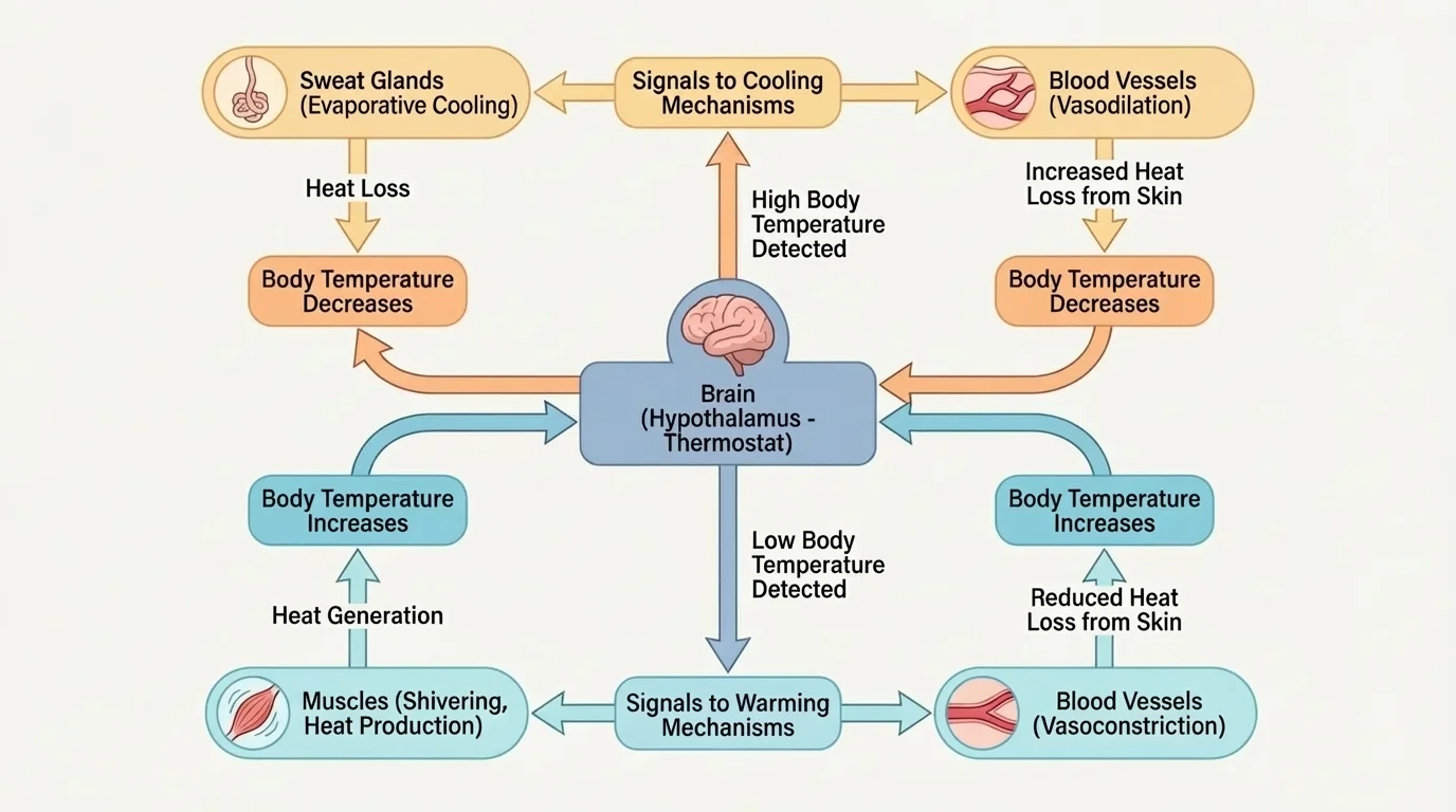
This is an example of a feedback loop, in which the output of a process influences the process itself. In negative feedback, the response reduces the original change. If temperature goes up, the response helps bring it down. If blood glucose rises after a meal, hormones help lower it toward normal range.
The endocrine system also contributes to regulation. Hormones travel through the blood and act on target cells. This means the endocrine system depends on the circulatory system for transport. The nervous and endocrine systems often work together: one sends rapid electrical signals, the other sends chemical messages that may act more slowly but last longer.
Regulation depends on communication. A body system cannot maintain stable conditions if receptors fail to detect change, control centers fail to process information, or effectors fail to respond. Homeostasis is not a property of one organ. It emerges from interactions among sensors, signaling pathways, tissues, and organs.
That is why models of homeostasis must show direction of information flow as well as movement of matter. In [Figure 4], arrows are not just decorative. They represent signals and responses that connect components into a working regulatory system.
A major reason to study system relationships is that problems rarely stay isolated. When one component of a biological system is damaged, the effects can spread. In anemia, for example, the blood has reduced capacity to carry oxygen. The lungs may function normally, but tissues still receive less oxygen, causing fatigue and weakness.
Kidney failure provides another example. The kidneys help regulate water balance, ion concentrations, and waste removal. If they fail, the composition of blood changes. This affects the circulatory system, nervous system, and many cellular processes. A model of kidney disease therefore needs to include more than the kidneys themselves.
Damage to neurons can also affect muscles and organs that depend on nerve signals. If motor neurons are impaired, muscles may not contract effectively. If sensory pathways are damaged, the body may fail to detect changes in the environment or within itself. The lesson is consistent across examples: function depends on connected parts.
Case study: dehydration during intense exercise
A student-athlete practices outdoors on a hot day and loses a large amount of water through sweat.
Step 1: Water loss reduces blood plasma volume.
Step 2: Reduced plasma volume makes it harder for the circulatory system to maintain efficient transport.
Step 3: Less effective circulation can reduce heat transfer to the skin and reduce oxygen delivery to muscles.
Step 4: Performance drops, body temperature may rise, and homeostatic regulation becomes more difficult.
This shows how the integumentary, circulatory, muscular, and nervous systems are linked during exercise.
Even at the molecular level, small changes can lead to system-wide consequences. If membrane proteins that move ions across cell membranes malfunction, cells may not maintain proper electrical activity or water balance. The problem begins with molecules but can affect tissues, organs, and the entire organism.
To build a useful model, start by asking a clear question. Are you trying to explain how nutrients reach cells, how a signal causes a response, or how a disease disrupts normal function? The question determines which components belong in the model.
Next, identify the major parts and the relationships among them. In a systems model, arrows often represent movement of matter, transfer of energy, or flow of information. Labels should be specific. Instead of drawing a general line from lung to body, a stronger model shows oxygen entering blood at alveoli, transport by circulation, and diffusion into cells.
| Model feature | What it should show | Why it matters |
|---|---|---|
| Components | Relevant structures such as organs, tissues, cells, or molecules | Identifies the parts involved in the function |
| Connections | Arrows or links between parts | Shows interaction, transport, or signaling |
| Evidence | Observed data, images, experiments, or measurements | Makes the model scientifically credible |
| Scale | The level of organization being represented | Prevents confusion between cell-level and organ-level events |
| Limitations | What the model leaves out | Helps users interpret it accurately |
Table 1. Features that make a biological model useful for explaining relationships within and between systems.
Evidence can also be used to revise models. Suppose data show that a patient's blood oxygen level is normal but muscles still fatigue quickly. That suggests the problem may not be lung function. The model might need to include cellular energy pathways, blood flow distribution, or muscle tissue damage. In science, models improve when new evidence appears.
Structure and function are closely linked in biology. Thin membranes, branching vessels, folded surfaces, and specialized cells are not random details. They are evidence for how a component contributes to the system's overall function.
A strong model is therefore not simply a map of parts. It is an explanation of how interactions produce a result. That idea applies whether the model focuses on one organ system or on relationships between multiple systems.
Doctors use system models when diagnosing disease. Shortness of breath may involve lungs, heart, blood chemistry, or even nervous system control of breathing. Athletes and trainers rely on models of oxygen delivery, hydration, and muscle energy use to improve performance safely. Biomedical engineers design artificial organs and medical devices by studying how biological components interact under normal and abnormal conditions.
Researchers also use models to predict responses. For instance, they can model how changes in ventilation affect blood gases, or how hormone levels influence blood glucose over time. Public health experts use models to understand how diseases that damage one system, such as respiratory infections, can have effects across the whole body.
"Nothing in biology makes sense except in the light of relationships."
— Adapted from a core idea in biological thinking
When you look at biology through models, the body stops seeming like a list of organs to memorize. It becomes a network of interacting systems, each dependent on the others, and our understanding of those systems is shaped by evidence from real observations. That is the heart of systems thinking in biology.